5YJ0

Mouse Cereblon thalidomide binding domain complexed with S-form thalidomide


Experimental Data Snapshot

  • Method: X-RAY DIFFRACTION
  • Resolution: 1.80 Å
  • R-Value Free: 0.204 
  • R-Value Work: 0.189 
  • R-Value Observed: 0.190 

wwPDB Validation   3D Report Full Report


Ligand Structure Quality Assessment 


This is version 1.2 of the entry. See complete history


Literature

Structural basis of thalidomide enantiomer binding to cereblon

Mori, T.Ito, T.Liu, S.Ando, H.Sakamoto, S.Yamaguchi, Y.Tokunaga, E.Shibata, N.Handa, H.Hakoshima, T.

(2018) Sci Rep 8: 1294-1294

  • DOI: https://doi.org/10.1038/s41598-018-19202-7
  • Primary Citation of Related Structures:  
    5YIZ, 5YJ0, 5YJ1

  • PubMed Abstract: 

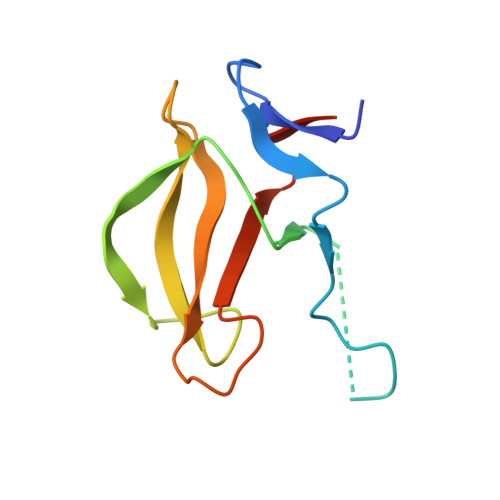
    Thalidomide possesses two optical isomers which have been reported to exhibit different pharmacological and toxicological activities. However, the precise mechanism by which the two isomers exert their different activities remains poorly understood. Here, we present structural and biochemical studies of (S)- and (R)-enantiomers bound to the primary target of thalidomide, cereblon (CRBN). Our biochemical studies employed deuterium-substituted thalidomides to suppress optical isomer conversion, and established that the (S)-enantiomer exhibited ~10-fold stronger binding to CRBN and inhibition of self-ubiquitylation compared to the (R)-enantiomer. The crystal structures of the thalidomide-binding domain of CRBN bound to each enantiomer show that both enantiomers bind the tri-Trp pocket, although the bound form of the (S)-enantiomer exhibited a more relaxed glutarimide ring conformation. The (S)-enantiomer induced greater teratogenic effects on fins of zebrafish compared to the (R)-enantiomer. This study has established a mechanism by which thalidomide exerts its effects in a stereospecific manner at the atomic level.


  • Organizational Affiliation

    Structural Biology Laboratory, Nara Institute of Science and Technology, 8916-5 Takayama, Ikoma, Nara, 630-0192, Japan.


Macromolecules
Find similar proteins by:  (by identity cutoff)  |  3D Structure
Entity ID: 1
MoleculeChains Sequence LengthOrganismDetailsImage
Protein cereblon111Mus musculusMutation(s): 0 
Gene Names: Crbn
UniProt & NIH Common Fund Data Resources
Find proteins for Q8C7D2 (Mus musculus)
Explore Q8C7D2 
Go to UniProtKB:  Q8C7D2
IMPC:  MGI:1913277
Entity Groups  
Sequence Clusters30% Identity50% Identity70% Identity90% Identity95% Identity100% Identity
UniProt GroupQ8C7D2
Sequence Annotations
Expand
  • Reference Sequence
Small Molecules
Ligands 3 Unique
IDChains Name / Formula / InChI Key2D Diagram3D Interactions
EF2
Query on EF2

Download Ideal Coordinates CCD File 
BA [auth J]
CB [auth e]
GA [auth M]
HB [auth h]
KA [auth P]
BA [auth J],
CB [auth e],
GA [auth M],
HB [auth h],
KA [auth P],
KB [auth k],
NA [auth S],
OB [auth n],
Q [auth A],
RA [auth V],
TB [auth q],
U [auth D],
WA [auth Y],
WB [auth t],
Y [auth G],
ZA [auth b]
S-Thalidomide
C13 H10 N2 O4
UEJJHQNACJXSKW-VIFPVBQESA-N
SO4
Query on SO4

Download Ideal Coordinates CCD File 
AA [auth G]
BB [auth b]
DA [auth J]
EA [auth J]
EB [auth e]
AA [auth G],
BB [auth b],
DA [auth J],
EA [auth J],
EB [auth e],
FA [auth J],
FB [auth e],
GB [auth e],
IA [auth M],
JA [auth M],
JB [auth h],
MA [auth P],
MB [auth k],
NB [auth k],
PA [auth S],
QA [auth S],
QB [auth n],
RB [auth n],
S [auth A],
SB [auth n],
T [auth A],
TA [auth V],
UA [auth V],
VA [auth V],
VB [auth q],
W [auth D],
X [auth D],
YA [auth Y],
YB [auth t],
ZB [auth t]
SULFATE ION
O4 S
QAOWNCQODCNURD-UHFFFAOYSA-L
ZN
Query on ZN

Download Ideal Coordinates CCD File 
AB [auth b]
CA [auth J]
DB [auth e]
HA [auth M]
IB [auth h]
AB [auth b],
CA [auth J],
DB [auth e],
HA [auth M],
IB [auth h],
LA [auth P],
LB [auth k],
OA [auth S],
PB [auth n],
R [auth A],
SA [auth V],
UB [auth q],
V [auth D],
XA [auth Y],
XB [auth t],
Z [auth G]
ZINC ION
Zn
PTFCDOFLOPIGGS-UHFFFAOYSA-N
Binding Affinity Annotations 
IDSourceBinding Affinity
EF2 BindingDB:  5YJ0 Ki: min: 8510, max: 2.27e+4 (nM) from 2 assay(s)
IC50: min: 268, max: 2.99e+4 (nM) from 5 assay(s)
Binding MOAD:  5YJ0 Kd: 5700 (nM) from 1 assay(s)
Experimental Data & Validation

Experimental Data

  • Method: X-RAY DIFFRACTION
  • Resolution: 1.80 Å
  • R-Value Free: 0.204 
  • R-Value Work: 0.189 
  • R-Value Observed: 0.190 
  • Space Group: H 3
Unit Cell:
Length ( Å )Angle ( ˚ )
a = 201.897α = 90
b = 201.897β = 90
c = 123.614γ = 120
Software Package:
Software NamePurpose
REFMACrefinement
HKL-2000data reduction
HKL-2000data scaling
PHASERphasing

Structure Validation

View Full Validation Report


Ligand Structure Quality Assessment 


Entry History 

Deposition Data

Revision History  (Full details and data files)

  • Version 1.0: 2018-02-07
    Type: Initial release
  • Version 1.1: 2018-02-14
    Changes: Structure summary
  • Version 1.2: 2023-11-22
    Changes: Data collection, Database references, Refinement description