5YIS

Crystal Structure of AnkB LIR/LC3B complex


Experimental Data Snapshot

  • Method: X-RAY DIFFRACTION
  • Resolution: 2.20 Å
  • R-Value Free: 0.250 
  • R-Value Work: 0.197 
  • R-Value Observed: 0.200 

wwPDB Validation   3D Report Full Report


This is version 1.3 of the entry. See complete history


Literature

Potent and specific Atg8-targeting autophagy inhibitory peptides from giant ankyrins.

Li, J.Zhu, R.Chen, K.Zheng, H.Zhao, H.Yuan, C.Zhang, H.Wang, C.Zhang, M.

(2018) Nat Chem Biol 14: 778-787

  • DOI: https://doi.org/10.1038/s41589-018-0082-8
  • Primary Citation of Related Structures:  
    5YIP, 5YIQ, 5YIR, 5YIS

  • PubMed Abstract: 

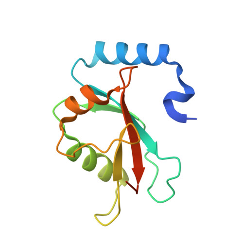
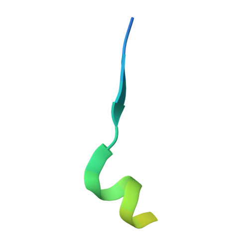
    The mammalian Atg8 family proteins are central drivers of autophagy and contain six members, classified into the LC3 and GABARAP subfamilies. Due to their high sequence similarity and consequent functional overlaps, it is difficult to delineate specific functions of Atg8 proteins in autophagy. Here we discover a super-strong GABARAP-selective inhibitory peptide harbored in 270/480 kDa ankyrin-G and a super-potent pan-Atg8 inhibitory peptide from 440 kDa ankyrin-B. Structural studies elucidate the mechanism governing the Atg8 binding potency and selectivity of the peptides, reveal a general Atg8-binding sequence motif, and allow development of a more GABARAP-selective inhibitory peptide. These peptides effectively blocked autophagy when expressed in cultured cells. Expression of these ankyrin-derived peptides in Caenorhabditis elegans also inhibited autophagy, causing accumulation of the p62 homolog SQST-1, delayed development and shortened life span. Thus, these genetically encodable autophagy inhibitory peptides can be used to occlude autophagy spatiotemporally in living animals.


  • Organizational Affiliation

    Division of Life Science, State Key Laboratory of Molecular Neuroscience, Hong Kong University of Science and Technology, Clear Water Bay, Kowloon, Hong Kong, China.


Macromolecules
Find similar proteins by:  (by identity cutoff)  |  3D Structure
Entity ID: 1
MoleculeChains Sequence LengthOrganismDetailsImage
Microtubule-associated proteins 1A/1B light chain 3B
A, B
125Mus musculusMutation(s): 0 
Gene Names: Map1lc3bMap1alc3Map1lc3
UniProt
Find proteins for Q9CQV6 (Mus musculus)
Explore Q9CQV6 
Go to UniProtKB:  Q9CQV6
Entity Groups  
Sequence Clusters30% Identity50% Identity70% Identity90% Identity95% Identity100% Identity
UniProt GroupQ9CQV6
Sequence Annotations
Expand
  • Reference Sequence
Find similar proteins by:  (by identity cutoff)  |  3D Structure
Entity ID: 2
MoleculeChains Sequence LengthOrganismDetailsImage
Ankyrin-2C [auth D],
D [auth C]
27Homo sapiensMutation(s): 0 
Gene Names: ANK2
UniProt & NIH Common Fund Data Resources
Find proteins for Q01484 (Homo sapiens)
Explore Q01484 
Go to UniProtKB:  Q01484
PHAROS:  Q01484
GTEx:  ENSG00000145362 
Entity Groups  
Sequence Clusters30% Identity50% Identity70% Identity90% Identity95% Identity100% Identity
UniProt GroupQ01484
Sequence Annotations
Expand
  • Reference Sequence
Experimental Data & Validation

Experimental Data

  • Method: X-RAY DIFFRACTION
  • Resolution: 2.20 Å
  • R-Value Free: 0.250 
  • R-Value Work: 0.197 
  • R-Value Observed: 0.200 
  • Space Group: P 21 21 21
Unit Cell:
Length ( Å )Angle ( ˚ )
a = 61.802α = 90
b = 66.866β = 90
c = 74.148γ = 90
Software Package:
Software NamePurpose
DENZOdata collection
SCALEPACKdata scaling
PHENIXrefinement
PHASERphasing
PDB_EXTRACTdata extraction

Structure Validation

View Full Validation Report


Entry History & Funding Information

Deposition Data


Funding OrganizationLocationGrant Number
Research Grants Council (RGC)Hong KongAoE-M09-12

Revision History  (Full details and data files)

  • Version 1.0: 2018-05-23
    Type: Initial release
  • Version 1.1: 2018-07-04
    Changes: Data collection, Database references
  • Version 1.2: 2018-08-01
    Changes: Data collection, Database references
  • Version 1.3: 2023-11-22
    Changes: Data collection, Database references, Refinement description