5YII

Crystal Structure of 45 amino acid deleted from N-terminal of Phosphoserine Aminotransferase (PSAT) of Entamoeba histolytica


Experimental Data Snapshot

  • Method: X-RAY DIFFRACTION
  • Resolution: 1.80 Å
  • R-Value Free: 0.269 
  • R-Value Work: 0.229 
  • R-Value Observed: 0.231 

wwPDB Validation   3D Report Full Report


This is version 1.2 of the entry. See complete history


Literature

N-terminal residues are crucial for quaternary structure and active site conformation for the phosphoserine aminotransferase from enteric human parasite E. histolytica.

Singh, R.K.Tomar, P.Dharavath, S.Kumar, S.Gourinath, S.

(2019) Int J Biol Macromol 132: 1012-1023

  • DOI: https://doi.org/10.1016/j.ijbiomac.2019.04.027
  • Primary Citation of Related Structures:  
    5YB0, 5YD2, 5YII

  • PubMed Abstract: 

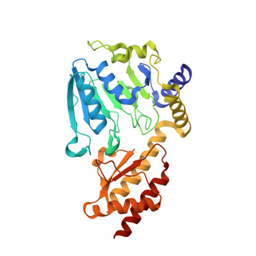
    Phosphoserine aminotransferase (PSAT) is a pyridoxal-5'phosphate (PLP)-dependent enzyme that catalyzes the second reversible step in the phosphoserine biosynthetic pathway producing serine. The crystal structure of E. histolytica PSAT (EhPSAT) complexed with PLP was elucidated at 3.0 Å resolution and the structures of its mutants, EhPSAT_Δ45 and EhPSAT_Δ4, at 1.8 and 2.4 Å resolution respectively. Deletion of 45 N-terminal residues (EhPSAT_Δ45) resulted in an inactive protein, the structure showed a dimeric arrangement drastically different from that of the wild-type protein, with the two monomers translated and rotated by almost 180° with respect to each other; causing a rearrangement of the active site to which PLP was unable to bind. Deletion of first N-terminal 15 (EhPSAT_Δ15) and four 11th to 14th residues (EhPSAT_Δ4) yielded up to 98% and 90% decrease in the activity respectively. Absence of aldimine linkage between PLP-Lys in the crystal structure of EhPSAT_Δ4 mutant explains for such decrease in activity and describes the importance of these N-terminal residues. Furthermore, a halide-binding site was found in close proximity to the active site. A stretch of six amino acids (146-NNTIYG-151) only conserved in the Entamoeba genus, contributes to halide binding may explain that the halide inhibition could be specific to Entamoeba.


  • Organizational Affiliation

    Structural Biology Laboratory, School of Life Sciences, Jawaharlal Nehru University, New Delhi 110067, India.


Macromolecules
Find similar proteins by:  (by identity cutoff)  |  3D Structure
Entity ID: 1
MoleculeChains Sequence LengthOrganismDetailsImage
Phosphoserine aminotransferase313Entamoeba histolyticaMutation(s): 0 
Gene Names: EhPSATEHI_026360
EC: 2.6.1.52
UniProt
Find proteins for Q60I38 (Entamoeba histolytica)
Explore Q60I38 
Go to UniProtKB:  Q60I38
Entity Groups  
Sequence Clusters30% Identity50% Identity70% Identity90% Identity95% Identity100% Identity
UniProt GroupQ60I38
Sequence Annotations
Expand
  • Reference Sequence
Small Molecules
Ligands 1 Unique
IDChains Name / Formula / InChI Key2D Diagram3D Interactions
SCN
Query on SCN

Download Ideal Coordinates CCD File 
B [auth A]THIOCYANATE ION
C N S
ZMZDMBWJUHKJPS-UHFFFAOYSA-M
Experimental Data & Validation

Experimental Data

  • Method: X-RAY DIFFRACTION
  • Resolution: 1.80 Å
  • R-Value Free: 0.269 
  • R-Value Work: 0.229 
  • R-Value Observed: 0.231 
  • Space Group: C 2 2 21
Unit Cell:
Length ( Å )Angle ( ˚ )
a = 72.256α = 90
b = 103.044β = 90
c = 80.83γ = 90
Software Package:
Software NamePurpose
REFMACrefinement
HKL-2000data collection
HKL-2000data scaling
PHASERphasing
PDB_EXTRACTdata extraction
HKL-2000data reduction
DMphasing

Structure Validation

View Full Validation Report


Entry History 

Deposition Data

Revision History  (Full details and data files)

  • Version 1.0: 2018-10-24
    Type: Initial release
  • Version 1.1: 2019-07-10
    Changes: Data collection, Database references
  • Version 1.2: 2023-11-22
    Changes: Data collection, Database references, Refinement description