5Y86

Crystal structure of kinase


Experimental Data Snapshot

  • Method: X-RAY DIFFRACTION
  • Resolution: 1.90 Å
  • R-Value Free: 0.196 
  • R-Value Work: 0.167 
  • R-Value Observed: 0.168 

wwPDB Validation   3D Report Full Report


Ligand Structure Quality Assessment 


This is version 1.2 of the entry. See complete history


Literature

Crystal Structure of Human Dual-Specificity Tyrosine-Regulated Kinase 3 Reveals New Structural Features and Insights into its Auto-phosphorylation

Kim, K.Cha, J.S.Cho, Y.S.Kim, H.Chang, N.Kim, H.J.Cho, H.S.

(2018) J Mol Biol 430: 1521-1530

  • DOI: https://doi.org/10.1016/j.jmb.2018.04.001
  • Primary Citation of Related Structures:  
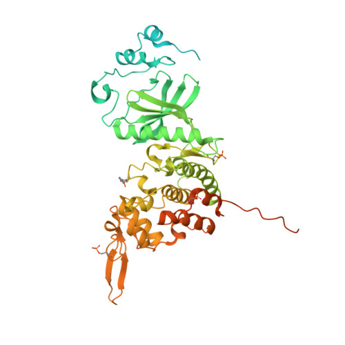
    5Y86

  • PubMed Abstract: 

    Dual-specificity tyrosine-regulated kinases (DYRKs) auto-phosphorylate a critical tyrosine residue in their activation loop and phosphorylate their substrate on serine and threonine residues. The auto-phosphorylation occurs intramolecularly and is a one-off event. DYRK3 is selectively expressed at a high level in hematopoietic cells and attenuates erythroblast development, leading to anemia. In the present study, we determined the crystal structure of the mature form of human DYRK3 in complex with harmine, an ATP competitive inhibitor. The crystal structure revealed a phosphorylation site, residue S350, whose phosphorylation increases the stability of DYRK3 and enhances its kinase activity. In addition, our structural and biochemical assays suggest that the N-terminal auto-phosphorylation accessory domain stabilizes the DYRK3 protein, followed by auto-phosphorylation of the tyrosine of the activation loop, which is important for kinase activity. Finally, our docking analysis provides information for the design of novel and potent therapeutics to treat anemia.


  • Organizational Affiliation

    Department of Systems Biology and Division of Life Sciences, Yonsei University, 50 Yonsei-ro, Seodaemun-gu, Seoul 03722, Republic of Korea.


Macromolecules
Find similar proteins by:  (by identity cutoff)  |  3D Structure
Entity ID: 1
MoleculeChains Sequence LengthOrganismDetailsImage
Dual specificity tyrosine-phosphorylation-regulated kinase 3588Homo sapiensMutation(s): 0 
Gene Names: DYRK3
EC: 2.7.12.1
UniProt & NIH Common Fund Data Resources
Find proteins for O43781 (Homo sapiens)
Explore O43781 
Go to UniProtKB:  O43781
PHAROS:  O43781
GTEx:  ENSG00000143479 
Entity Groups  
Sequence Clusters30% Identity50% Identity70% Identity90% Identity95% Identity100% Identity
UniProt GroupO43781
Sequence Annotations
Expand
  • Reference Sequence
Small Molecules
Ligands 4 Unique
IDChains Name / Formula / InChI Key2D Diagram3D Interactions
HRM (Subject of Investigation/LOI)
Query on HRM

Download Ideal Coordinates CCD File 
B [auth A]7-METHOXY-1-METHYL-9H-BETA-CARBOLINE
C13 H12 N2 O
BXNJHAXVSOCGBA-UHFFFAOYSA-N
PO4
Query on PO4

Download Ideal Coordinates CCD File 
C [auth A],
D [auth A]
PHOSPHATE ION
O4 P
NBIIXXVUZAFLBC-UHFFFAOYSA-K
GOL
Query on GOL

Download Ideal Coordinates CCD File 
M [auth A]GLYCEROL
C3 H8 O3
PEDCQBHIVMGVHV-UHFFFAOYSA-N
EDO
Query on EDO

Download Ideal Coordinates CCD File 
E [auth A]
F [auth A]
G [auth A]
H [auth A]
I [auth A]
E [auth A],
F [auth A],
G [auth A],
H [auth A],
I [auth A],
J [auth A],
K [auth A],
L [auth A]
1,2-ETHANEDIOL
C2 H6 O2
LYCAIKOWRPUZTN-UHFFFAOYSA-N
Modified Residues  3 Unique
IDChains TypeFormula2D DiagramParent
PTR
Query on PTR
A
L-PEPTIDE LINKINGC9 H12 N O6 PTYR
SEP
Query on SEP
A
L-PEPTIDE LINKINGC3 H8 N O6 PSER
TPO
Query on TPO
A
L-PEPTIDE LINKINGC4 H10 N O6 PTHR
Binding Affinity Annotations 
IDSourceBinding Affinity
HRM BindingDB:  5Y86 IC50: min: 210, max: 800 (nM) from 3 assay(s)
Binding MOAD:  5Y86 IC50: 800 (nM) from 1 assay(s)
Experimental Data & Validation

Experimental Data

  • Method: X-RAY DIFFRACTION
  • Resolution: 1.90 Å
  • R-Value Free: 0.196 
  • R-Value Work: 0.167 
  • R-Value Observed: 0.168 
  • Space Group: P 2 21 21
Unit Cell:
Length ( Å )Angle ( ˚ )
a = 43α = 90
b = 76.52β = 90
c = 148.47γ = 90
Software Package:
Software NamePurpose
REFMACrefinement
HKL-3000data collection
HKL-3000data scaling
MOLREPphasing

Structure Validation

View Full Validation Report


Ligand Structure Quality Assessment 


Entry History 

Deposition Data

Revision History  (Full details and data files)

  • Version 1.0: 2018-05-02
    Type: Initial release
  • Version 1.1: 2018-05-30
    Changes: Data collection, Database references
  • Version 1.2: 2023-11-22
    Changes: Data collection, Database references, Refinement description, Structure summary