5Y6Q

Crystal structure of an aldehyde oxidase from Methylobacillus sp. KY4400


Experimental Data Snapshot

  • Method: X-RAY DIFFRACTION
  • Resolution: 2.50 Å
  • R-Value Free: 0.233 
  • R-Value Work: 0.178 
  • R-Value Observed: 0.181 

wwPDB Validation   3D Report Full Report


Ligand Structure Quality Assessment 


This is version 1.2 of the entry. See complete history


Literature

Crystal structure of an aldehyde oxidase from Methylobacillus sp. KY4400.

Uchida, H.Mikami, B.Yamane-Tanabe, A.Ito, A.Hirano, K.Oki, M.

(2018) J Biochem 163: 321-328

  • DOI: https://doi.org/10.1093/jb/mvy004
  • Primary Citation of Related Structures:  
    5Y6Q

  • PubMed Abstract: 

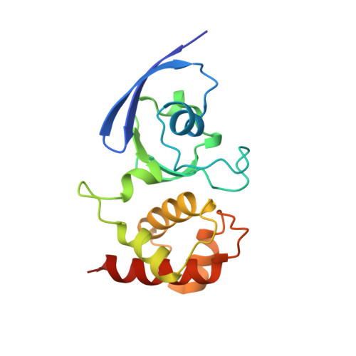
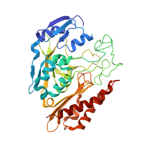
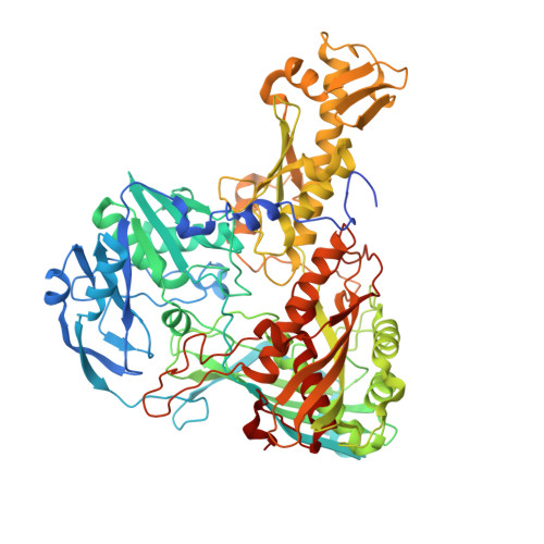
    Hetero-trimeric aldehyde oxidases of bacterial origin, which use O2 to catalyse the oxidation of various aldehydes but not those of aromatic N-heterocycles, belong to the xanthine oxidase family. In the present study, the crystal structure of a recombinant aldehyde oxidase from Methylobacillus sp. KY4400 (Mb-AOX) was determined at 2.5 Å resolution. The structures of its subunits resemble those of the corresponding subunits or domains of other structurally characterised enzymes belonging to the family, and include a [4Fe-4 S] cluster in the medium subunit like that found in Escherichia coli periplasmic aldehyde oxidoreductase (EP-AOR). A funnel leading to the si-face of the isoalloxazine ring of FAD, which is narrower than those in mouse liver AOX3 and human AOX1, is also present and it is even narrower than that in EP-AOR. The environment surrounding the ring in Mb-AOX and EP-AOR is subtly different, which might account for their different abilities to use O2. A remarkable characteristic of the Mo catalytic centre in Mb-AOX is a tryptophan situated near the centre instead of the alanine present in other xanthine oxidase family members. The tryptophan residue together with other residue differences might play an important role in binding to aldehydes such as n-heptylaldehyde in Mb-AOX.


  • Organizational Affiliation

    Department of Applied Chemistry and Biotechnology, Graduate School of Engineering, University of Fukui, 9-1, Bunkyo 3-Chome, Fukui 910-8507, Japan.


Macromolecules
Find similar proteins by:  (by identity cutoff)  |  3D Structure
Entity ID: 1
MoleculeChains Sequence LengthOrganismDetailsImage
Aldehyde oxidase small subunit162Methylobacillus sp. KY4400Mutation(s): 0 
Gene Names: aoms
UniProt
Find proteins for Q84IY0 (Methylobacillus sp. KY4400)
Explore Q84IY0 
Go to UniProtKB:  Q84IY0
Entity Groups  
Sequence Clusters30% Identity50% Identity70% Identity90% Identity95% Identity100% Identity
UniProt GroupQ84IY0
Sequence Annotations
Expand
  • Reference Sequence
Find similar proteins by:  (by identity cutoff)  |  3D Structure
Entity ID: 2
MoleculeChains Sequence LengthOrganismDetailsImage
Aldehyde oxidase medium subunit330Methylobacillus sp. KY4400Mutation(s): 0 
Gene Names: aomm
UniProt
Find proteins for Q84IX9 (Methylobacillus sp. KY4400)
Explore Q84IX9 
Go to UniProtKB:  Q84IX9
Entity Groups  
Sequence Clusters30% Identity50% Identity70% Identity90% Identity95% Identity100% Identity
UniProt GroupQ84IX9
Sequence Annotations
Expand
  • Reference Sequence
Find similar proteins by:  (by identity cutoff)  |  3D Structure
Entity ID: 3
MoleculeChains Sequence LengthOrganismDetailsImage
Aldehyde oxidase large subunit775Methylobacillus sp. KY4400Mutation(s): 0 
Gene Names: aoml
UniProt
Find proteins for Q84IX8 (Methylobacillus sp. KY4400)
Explore Q84IX8 
Go to UniProtKB:  Q84IX8
Entity Groups  
Sequence Clusters30% Identity50% Identity70% Identity90% Identity95% Identity100% Identity
UniProt GroupQ84IX8
Sequence Annotations
Expand
  • Reference Sequence
Small Molecules
Ligands 8 Unique
IDChains Name / Formula / InChI Key2D Diagram3D Interactions
FAD
Query on FAD

Download Ideal Coordinates CCD File 
G [auth B]FLAVIN-ADENINE DINUCLEOTIDE
C27 H33 N9 O15 P2
VWWQXMAJTJZDQX-UYBVJOGSSA-N
MCN
Query on MCN

Download Ideal Coordinates CCD File 
O [auth C]PTERIN CYTOSINE DINUCLEOTIDE
C19 H22 N8 O13 P2 S2
RBWYFPNWTRZKKZ-LOIMWUFNSA-N
SF4
Query on SF4

Download Ideal Coordinates CCD File 
H [auth B]IRON/SULFUR CLUSTER
Fe4 S4
LJBDFODJNLIPKO-UHFFFAOYSA-N
FES
Query on FES

Download Ideal Coordinates CCD File 
D [auth A],
E [auth A]
FE2/S2 (INORGANIC) CLUSTER
Fe2 S2
NIXDOXVAJZFRNF-UHFFFAOYSA-N
MOS
Query on MOS

Download Ideal Coordinates CCD File 
N [auth C]DIOXOTHIOMOLYBDENUM(VI) ION
H Mo O2 S
BDSRWPHSAKXXRG-UHFFFAOYSA-M
SO4
Query on SO4

Download Ideal Coordinates CCD File 
F [auth A]
I [auth B]
J [auth B]
K [auth B]
P [auth C]
F [auth A],
I [auth B],
J [auth B],
K [auth B],
P [auth C],
Q [auth C],
R [auth C],
S [auth C],
T [auth C],
U [auth C],
V [auth C],
W [auth C]
SULFATE ION
O4 S
QAOWNCQODCNURD-UHFFFAOYSA-L
GOL
Query on GOL

Download Ideal Coordinates CCD File 
AA [auth C]
L [auth B]
M [auth B]
X [auth C]
Y [auth C]
AA [auth C],
L [auth B],
M [auth B],
X [auth C],
Y [auth C],
Z [auth C]
GLYCEROL
C3 H8 O3
PEDCQBHIVMGVHV-UHFFFAOYSA-N
IPA
Query on IPA

Download Ideal Coordinates CCD File 
BA [auth C],
CA [auth C]
ISOPROPYL ALCOHOL
C3 H8 O
KFZMGEQAYNKOFK-UHFFFAOYSA-N
Experimental Data & Validation

Experimental Data

  • Method: X-RAY DIFFRACTION
  • Resolution: 2.50 Å
  • R-Value Free: 0.233 
  • R-Value Work: 0.178 
  • R-Value Observed: 0.181 
  • Space Group: P 41 21 2
Unit Cell:
Length ( Å )Angle ( ˚ )
a = 77.972α = 90
b = 77.972β = 90
c = 400.34γ = 90
Software Package:
Software NamePurpose
PHENIXrefinement
DENZOdata reduction
SCALEPACKdata scaling
MOLREPphasing

Structure Validation

View Full Validation Report


Ligand Structure Quality Assessment 


Entry History & Funding Information

Deposition Data


Funding OrganizationLocationGrant Number
Japan--

Revision History  (Full details and data files)

  • Version 1.0: 2018-01-24
    Type: Initial release
  • Version 1.1: 2018-04-11
    Changes: Data collection, Database references
  • Version 1.2: 2023-11-22
    Changes: Data collection, Database references, Refinement description