


Crystal structure of Pseudomonas aeruginosa HmgR
Gu, T., Ji, Q., Liang, H.To be published.
Experimental Data Snapshot
wwPDB Validation 3D Report Full Report

Entity ID: 1 | |||||
|---|---|---|---|---|---|
| Molecule | Chains | Sequence Length | Organism | Details | Image |
| Transcriptional regulator KdgR | 267 | Pseudomonas aeruginosa | Mutation(s): 0 Gene Names: kdgR_2, AOY09_03044, PAERUG_P32_London_17_VIM_2_10_11_04319 | 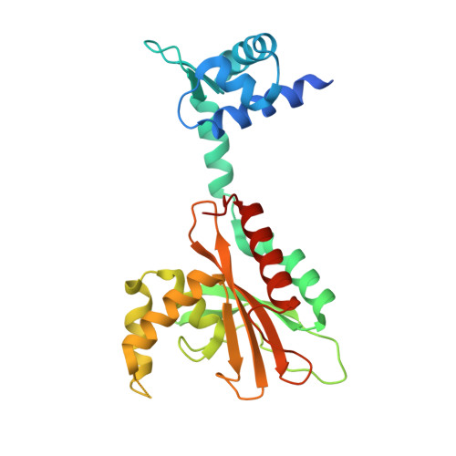 | |
UniProt | |||||
Find proteins for Q9I2A1 (Pseudomonas aeruginosa (strain ATCC 15692 / DSM 22644 / CIP 104116 / JCM 14847 / LMG 12228 / 1C / PRS 101 / PAO1)) Explore Q9I2A1 Go to UniProtKB: Q9I2A1 | |||||
Entity Groups | |||||
| Sequence Clusters | 30% Identity50% Identity70% Identity90% Identity95% Identity100% Identity | ||||
| UniProt Group | Q9I2A1 | ||||
Sequence AnnotationsExpand | |||||
| |||||
| Length ( Å ) | Angle ( ˚ ) |
|---|---|
| a = 60.129 | α = 90 |
| b = 80.52 | β = 90 |
| c = 116.755 | γ = 90 |
| Software Name | Purpose |
|---|---|
| PHENIX | refinement |
| HKL-3000 | data reduction |
| HKL-3000 | data scaling |
| PHASER | phasing |
RCSB PDB (citation) is hosted by
RCSB PDB is a member of the

