5Y6F

Crystal structure of YcgR in complex with c-di-GMP from Escherichia coli


Experimental Data Snapshot

  • Method: X-RAY DIFFRACTION
  • Resolution: 2.30 Å
  • R-Value Free: 0.233 
  • R-Value Work: 0.183 
  • R-Value Observed: 0.185 

wwPDB Validation   3D Report Full Report


Ligand Structure Quality Assessment 


This is version 1.2 of the entry. See complete history


Literature

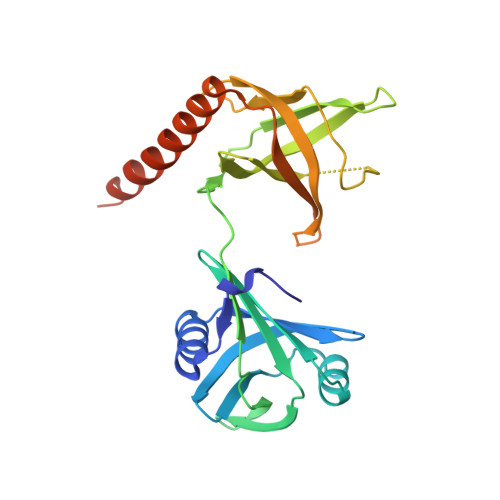
Structural insights into the mechanism of c-di-GMP-bound YcgR regulating flagellar motility inEscherichia coli.

Hou, Y.J.Yang, W.S.Hong, Y.Zhang, Y.Wang, D.C.Li, D.F.

(2020) J Biol Chem 295: 808-821

  • DOI: https://doi.org/10.1074/jbc.RA119.009739
  • Primary Citation of Related Structures:  
    5Y6F, 5Y6G, 5Y6H

  • PubMed Abstract: 

    The motile-sessile transition is critical for bacterial survival and growth. Cyclic-di-GMP (c-di-GMP) plays a central role in controlling this transition and regulating biofilm formation via various effectors. As an effector of c-di-GMP in Escherichia coli and related species, the PilZ domain-containing protein YcgR responds to elevated c-di-GMP concentrations and acts on the flagellar motor to suppress bacterial motility in a brakelike fashion, which promotes bacterial surface attachment. To date, several target proteins within the motor, MotA, FliG, and FliM, along with different regulatory mechanisms have been reported. However, how YcgR acts on these components remains unclear. Here, we report that activated YcgR stably binds to MotA at the MotA-FliG interface and thereby regulates bacterial swimming. Biochemical and structural analyses revealed that c-di-GMP rearranges the PilZ domain configuration, resulting in the formation of a MotA-binding patch consisting of an R XXX R motif and the C-tail helix α3. Moreover, we noted that a conserved region in the YcgR-N domain, which is independent of MotA interaction, is necessary for motility regulation. On the basis of these findings, we infer that the YcgR-N domain is required for activity on other motor proteins. We propose that activated YcgR appends to MotA via its PilZ domain and thereby interrupts the MotA-FliG interaction and simultaneously interacts with other motor proteins via its YcgR-N domain to inhibit flagellar motility. Our findings suggest that the mode of interaction between YcgR and motor proteins may be shared by other PilZ family proteins.


  • Organizational Affiliation

    National Laboratory of Biomacromolecules, CAS Center for Excellence in Biomacromolecules, Institute of Biophysics, Chinese Academy of Sciences, Beijing 100101, China.


Macromolecules
Find similar proteins by:  (by identity cutoff)  |  3D Structure
Entity ID: 1
MoleculeChains Sequence LengthOrganismDetailsImage
Flagellar brake protein YcgR257Escherichia coli K-12Mutation(s): 0 
Gene Names: ycgRb1194JW1183
UniProt
Find proteins for P76010 (Escherichia coli (strain K12))
Explore P76010 
Go to UniProtKB:  P76010
Entity Groups  
Sequence Clusters30% Identity50% Identity70% Identity90% Identity95% Identity100% Identity
UniProt GroupP76010
Sequence Annotations
Expand
  • Reference Sequence
Small Molecules
Ligands 2 Unique
IDChains Name / Formula / InChI Key2D Diagram3D Interactions
C2E (Subject of Investigation/LOI)
Query on C2E

Download Ideal Coordinates CCD File 
C [auth A],
D [auth A]
9,9'-[(2R,3R,3aS,5S,7aR,9R,10R,10aS,12S,14aR)-3,5,10,12-tetrahydroxy-5,12-dioxidooctahydro-2H,7H-difuro[3,2-d:3',2'-j][1,3,7,9,2,8]tetraoxadiphosphacyclododecine-2,9-diyl]bis(2-amino-1,9-dihydro-6H-purin-6-one)
C20 H24 N10 O14 P2
PKFDLKSEZWEFGL-MHARETSRSA-N
SO4
Query on SO4

Download Ideal Coordinates CCD File 
B [auth A]SULFATE ION
O4 S
QAOWNCQODCNURD-UHFFFAOYSA-L
Experimental Data & Validation

Experimental Data

  • Method: X-RAY DIFFRACTION
  • Resolution: 2.30 Å
  • R-Value Free: 0.233 
  • R-Value Work: 0.183 
  • R-Value Observed: 0.185 
  • Space Group: H 3
Unit Cell:
Length ( Å )Angle ( ˚ )
a = 93.98α = 90
b = 93.98β = 90
c = 109.64γ = 120
Software Package:
Software NamePurpose
PHENIXrefinement
iMOSFLMdata reduction
SCALAdata scaling
PHASERphasing

Structure Validation

View Full Validation Report


Ligand Structure Quality Assessment 


Entry History 

Deposition Data

Revision History  (Full details and data files)

  • Version 1.0: 2018-07-18
    Type: Initial release
  • Version 1.1: 2020-02-05
    Changes: Database references, Structure summary
  • Version 1.2: 2024-03-27
    Changes: Data collection, Database references