5Y5W

Crystal structure of human Spindlin1 in complex with a histone H4K20(me3) peptide


Experimental Data Snapshot

  • Method: X-RAY DIFFRACTION
  • Resolution: 3.30 Å
  • R-Value Free: 0.308 
  • R-Value Work: 0.219 
  • R-Value Observed: 0.224 

wwPDB Validation   3D Report Full Report


This is version 1.3 of the entry. See complete history


Literature

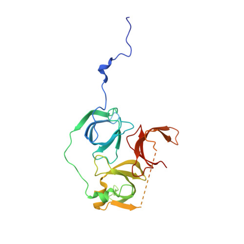
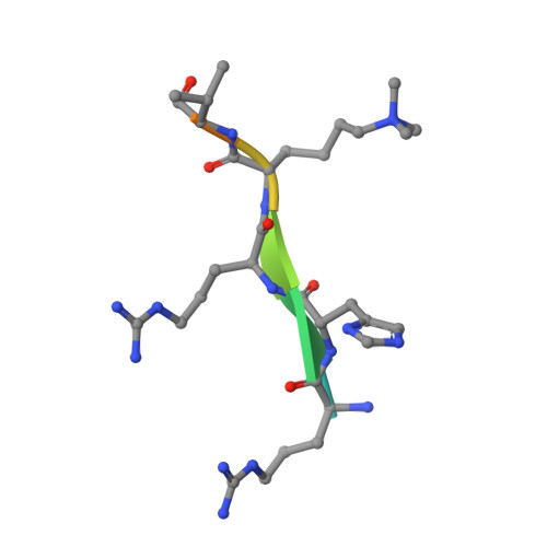
Spindlin-1 recognizes methylations of K20 and R23 of histone H4 tail

Wang, C.Zhan, L.Wu, M.Ma, R.Yao, J.Xiong, Y.Pan, Y.Guan, S.Zhang, X.Zang, J.

(2018) FEBS Lett 592: 4098-4110

  • DOI: https://doi.org/10.1002/1873-3468.13281
  • Primary Citation of Related Structures:  
    5Y5W

  • PubMed Abstract: 

    Using methods combining cross-linking, pull-down assays, and stable isotope labeling by amino acids in cell culture with mass spectrometry, we identified that the Tudor domain-containing protein Spindlin-1 recognizes trimethylation of histone H4 lysine 20 (H4K20me3). The binding affinity of Spindlin-1 to H4K20me3 is weaker than that to H3K4me3, indicating H4K20me3 as a secondary substrate for Spindlin-1. Structural studies of Spindlin-1 in complex with the H4K20me3 peptide indicate that Spindlin-1 attains a distinct binding mode for H4K20me3 recognition. Further biochemical analysis identified that Spindlin-1 also binds methylated R23 of H4, providing new clues for the function of Spindlin-1.


  • Organizational Affiliation

    Hefei National Laboratory for Physical Sciences at Microscale CAS Center for Excellence in Biomacromolecules, Collaborative Innovation Center of Chemistry for Life Sciences and School of Life Sciences, University of Science and Technology of China, Hefei, China.


Macromolecules
Find similar proteins by:  (by identity cutoff)  |  3D Structure
Entity ID: 1
MoleculeChains Sequence LengthOrganismDetailsImage
Spindlin-1
A, B, C, D
235Homo sapiensMutation(s): 0 
Gene Names: SPIN1OCRSPIN
UniProt & NIH Common Fund Data Resources
Find proteins for Q9Y657 (Homo sapiens)
Explore Q9Y657 
Go to UniProtKB:  Q9Y657
PHAROS:  Q9Y657
GTEx:  ENSG00000106723 
Entity Groups  
Sequence Clusters30% Identity50% Identity70% Identity90% Identity95% Identity100% Identity
UniProt GroupQ9Y657
Sequence Annotations
Expand
  • Reference Sequence

Find similar proteins by:  Sequence   |   3D Structure  

Entity ID: 2
MoleculeChains Sequence LengthOrganismDetailsImage
Histone peptide H4K20(me3)
E, F, G
9Homo sapiensMutation(s): 0 
Sequence Annotations
Expand
  • Reference Sequence
Small Molecules
Modified Residues  1 Unique
IDChains TypeFormula2D DiagramParent
M3L
Query on M3L
E, F, G
L-PEPTIDE LINKINGC9 H21 N2 O2LYS
Experimental Data & Validation

Experimental Data

  • Method: X-RAY DIFFRACTION
  • Resolution: 3.30 Å
  • R-Value Free: 0.308 
  • R-Value Work: 0.219 
  • R-Value Observed: 0.224 
  • Space Group: C 2 2 21
Unit Cell:
Length ( Å )Angle ( ˚ )
a = 88.769α = 90
b = 148.504β = 90
c = 169.212γ = 90
Software Package:
Software NamePurpose
REFMACrefinement
HKL-2000data reduction
HKL-2000data scaling
PHASERphasing

Structure Validation

View Full Validation Report


Entry History 

Deposition Data

Revision History  (Full details and data files)

  • Version 1.0: 2017-10-11
    Type: Initial release
  • Version 1.1: 2019-03-20
    Changes: Data collection, Database references, Structure summary
  • Version 1.2: 2019-03-27
    Changes: Data collection, Derived calculations
  • Version 1.3: 2023-11-22
    Changes: Data collection, Database references, Refinement description