5Y3T

Crystal structure of hetero-trimeric core of LUBAC: HOIP double-UBA complexed with HOIL-1L UBL and SHARPIN UBL


Experimental Data Snapshot

  • Method: X-RAY DIFFRACTION
  • Resolution: 2.40 Å
  • R-Value Free: 0.240 
  • R-Value Work: 0.186 
  • R-Value Observed: 0.190 

wwPDB Validation   3D Report Full Report


This is version 1.2 of the entry. See complete history


Literature

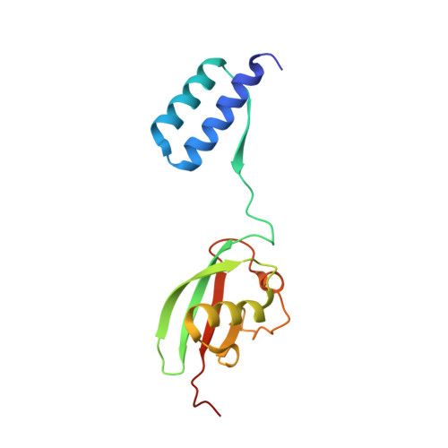
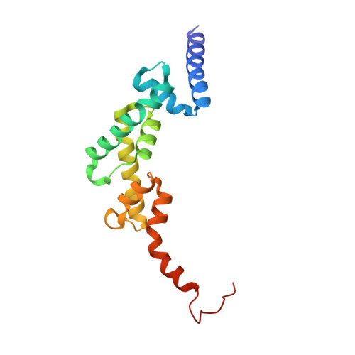
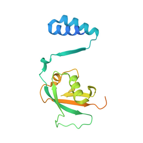
Cooperative Domain Formation by Homologous Motifs in HOIL-1L and SHARPIN Plays A Crucial Role in LUBAC Stabilization.

Fujita, H.Tokunaga, A.Shimizu, S.Whiting, A.L.Aguilar-Alonso, F.Takagi, K.Walinda, E.Sasaki, Y.Shimokawa, T.Mizushima, T.Ohki, I.Ariyoshi, M.Tochio, H.Bernal, F.Shirakawa, M.Iwai, K.

(2018) Cell Rep 23: 1192-1204

  • DOI: https://doi.org/10.1016/j.celrep.2018.03.112
  • Primary Citation of Related Structures:  
    5Y3T

  • PubMed Abstract: 

    The linear ubiquitin chain assembly complex (LUBAC) participates in inflammatory and oncogenic signaling by conjugating linear ubiquitin chains to target proteins. LUBAC consists of the catalytic HOIP subunit and two accessory subunits, HOIL-1L and SHARPIN. Interactions between the ubiquitin-associated (UBA) domains of HOIP and the ubiquitin-like (UBL) domains of two accessory subunits are involved in LUBAC stabilization, but the precise molecular mechanisms underlying the formation of stable trimeric LUBAC remain elusive. We solved the co-crystal structure of the binding regions of the trimeric LUBAC complex and found that LUBAC-tethering motifs (LTMs) located N terminally to the UBL domains of HOIL-1L and SHARPIN heterodimerize and fold into a single globular domain. This interaction is resistant to dissociation and plays a critical role in stabilizing trimeric LUBAC. Inhibition of LTM-mediated HOIL-1L/SHARPIN dimerization profoundly attenuated the function of LUBAC, suggesting LTM as a superior target of LUBAC destabilization for anticancer therapeutics.


  • Organizational Affiliation

    Department of Molecular and Cellular Physiology, Kyoto University School of Medicine, Kyoto 606-8501, Japan.


Macromolecules
Find similar proteins by:  (by identity cutoff)  |  3D Structure
Entity ID: 1
MoleculeChains Sequence LengthOrganismDetailsImage
RanBP-type and C3HC4-type zinc finger-containing protein 1140Mus musculusMutation(s): 0 
Gene Names: Rbck1RbckUbce7ip3Uip28
EC: 2.3.2.27
UniProt & NIH Common Fund Data Resources
Find proteins for Q9WUB0 (Mus musculus)
Explore Q9WUB0 
Go to UniProtKB:  Q9WUB0
IMPC:  MGI:1344372
Entity Groups  
Sequence Clusters30% Identity50% Identity70% Identity90% Identity95% Identity100% Identity
UniProt GroupQ9WUB0
Sequence Annotations
Expand
  • Reference Sequence
Find similar proteins by:  (by identity cutoff)  |  3D Structure
Entity ID: 2
MoleculeChains Sequence LengthOrganismDetailsImage
E3 ubiquitin-protein ligase RNF31158Mus musculusMutation(s): 0 
Gene Names: Rnf31Paul
EC: 2.3.2.27
UniProt & NIH Common Fund Data Resources
Find proteins for Q924T7 (Mus musculus)
Explore Q924T7 
Go to UniProtKB:  Q924T7
IMPC:  MGI:1934704
Entity Groups  
Sequence Clusters30% Identity50% Identity70% Identity90% Identity95% Identity100% Identity
UniProt GroupQ924T7
Sequence Annotations
Expand
  • Reference Sequence
Find similar proteins by:  (by identity cutoff)  |  3D Structure
Entity ID: 3
MoleculeChains Sequence LengthOrganismDetailsImage
Sharpin187Mus musculusMutation(s): 0 
Gene Names: SharpinCpdmSipl1
UniProt
Find proteins for Q91WA6 (Mus musculus)
Explore Q91WA6 
Go to UniProtKB:  Q91WA6
Entity Groups  
Sequence Clusters30% Identity50% Identity70% Identity90% Identity95% Identity100% Identity
UniProt GroupQ91WA6
Sequence Annotations
Expand
  • Reference Sequence
Small Molecules
Ligands 1 Unique
IDChains Name / Formula / InChI Key2D Diagram3D Interactions
GOL
Query on GOL

Download Ideal Coordinates CCD File 
D [auth B]GLYCEROL
C3 H8 O3
PEDCQBHIVMGVHV-UHFFFAOYSA-N
Experimental Data & Validation

Experimental Data

  • Method: X-RAY DIFFRACTION
  • Resolution: 2.40 Å
  • R-Value Free: 0.240 
  • R-Value Work: 0.186 
  • R-Value Observed: 0.190 
  • Space Group: C 1 2 1
Unit Cell:
Length ( Å )Angle ( ˚ )
a = 143.902α = 90
b = 59.732β = 97.42
c = 58.554γ = 90
Software Package:
Software NamePurpose
REFMACrefinement
HKL-2000data reduction
HKL-2000data scaling
MOLREPphasing

Structure Validation

View Full Validation Report


Entry History & Funding Information

Deposition Data


Funding OrganizationLocationGrant Number
Japan Society for the Promotion of ScienceJapanJP26119004
Japan Society for the Promotion of ScienceJapan15H01182

Revision History  (Full details and data files)

  • Version 1.0: 2018-05-02
    Type: Initial release
  • Version 1.1: 2019-12-25
    Changes: Database references
  • Version 1.2: 2023-11-22
    Changes: Data collection, Database references, Refinement description