5Y2B

Crystal structure of Ostrinia furnacalis Group II chitinase catalytic domain 1 in complex with HEPTA-N-ACETYLCHITOOCTAOSE (NAG)7


Experimental Data Snapshot

  • Method: X-RAY DIFFRACTION
  • Resolution: 2.20 Å
  • R-Value Free: 0.202 
  • R-Value Work: 0.153 

wwPDB Validation   3D Report Full Report


Ligand Structure Quality Assessment 


This is version 2.1 of the entry. See complete history


Literature

Structural analysis of group II chitinase (ChtII) catalysis completes the puzzle of chitin hydrolysis in insects

Chen, W.Qu, M.Zhou, Y.Yang, Q.

(2018) J Biol Chem 293: 2652-2660

  • DOI: https://doi.org/10.1074/jbc.RA117.000119
  • Primary Citation of Related Structures:  
    5Y29, 5Y2A, 5Y2B, 5Y2C

  • PubMed Abstract: 

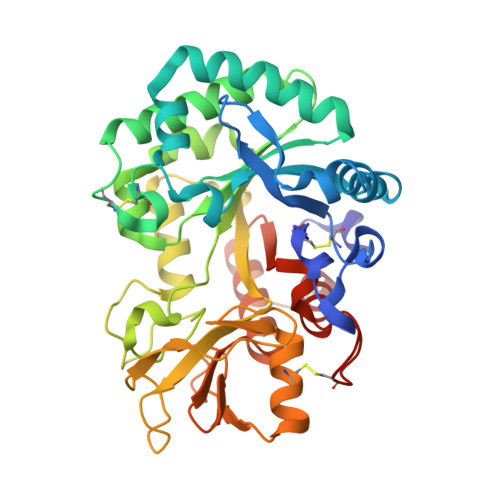
    Chitin is a linear homopolymer of N -acetyl-β-d-glucosamines and a major structural component of insect cuticles. Chitin hydrolysis involves glycoside hydrolase family 18 (GH18) chitinases. In insects, chitin hydrolysis is essential for periodic shedding of the old cuticle ecdysis and proceeds via a pathway different from that in the well studied bacterial chitinolytic system. Group II chitinase (ChtII) is a widespread chitinolytic enzyme in insects and contains the greatest number of catalytic domains and chitin-binding domains among chitinases. In Lepidopterans, ChtII and two other chitinases, ChtI and Chi-h, are essential for chitin hydrolysis. Although ChtI and Chi-h have been well studied, the role of ChtII remains elusive. Here, we investigated the structure and enzymology of Of ChtII, a ChtII derived from the insect pest Ostrinia furnacalis We present the crystal structures of two catalytically active domains of Of ChtII, Of ChtII-C1 and Of ChtII-C2, both in unliganded form and complexed with chitooligosaccharide substrates. We found that Of ChtII-C1 and Of ChtII-C2 both possess long, deep substrate-binding clefts with endochitinase activities. Of ChtII exhibited structural characteristics within the substrate-binding cleft similar to those in Of Chi-h and Of ChtI. However, Of ChtII lacked structural elements favoring substrate binding beyond the active sites, including an extra wall structure present in Of Chi-h. Nevertheless, the numerous domains in Of ChtII may compensate for this difference; a truncation containing one catalytic domain and three chitin-binding modules ( Of ChtII-B4C1) displayed activity toward insoluble polymeric substrates that was higher than those of Of Chi-h and Of ChtI. Our observations provide the last piece of the puzzle of chitin hydrolysis in insects.


  • Organizational Affiliation

    State Key Laboratory of Fine Chemical Engineering, School of Life Science and Biotechnology and School of Software, Dalian University of Technology, Dalian 116024, China.


Macromolecules
Find similar proteins by:  (by identity cutoff)  |  3D Structure
Entity ID: 1
MoleculeChains Sequence LengthOrganismDetailsImage
insect group II chitinase377Ostrinia furnacalisMutation(s): 0 
UniProt
Find proteins for A0A221ZS22 (Ostrinia furnacalis)
Explore A0A221ZS22 
Go to UniProtKB:  A0A221ZS22
Entity Groups  
Sequence Clusters30% Identity50% Identity70% Identity90% Identity95% Identity100% Identity
UniProt GroupA0A221ZS22
Sequence Annotations
Expand
  • Reference Sequence
Oligosaccharides

Help

Entity ID: 2
MoleculeChains Length2D Diagram Glycosylation3D Interactions
2-acetamido-2-deoxy-beta-D-glucopyranose-(1-4)-2-acetamido-2-deoxy-beta-D-glucopyranose-(1-4)-2-acetamido-2-deoxy-beta-D-glucopyranose-(1-4)-2-acetamido-2-deoxy-beta-D-glucopyranose-(1-4)-2-acetamido-2-deoxy-beta-D-glucopyranose-(1-4)-2-acetamido-2-deoxy-beta-D-glucopyranose-(1-4)-2-acetamido-2-deoxy-beta-D-glucopyranose
B
7N/A
Glycosylation Resources
GlyTouCan:  G15210HV
GlyCosmos:  G15210HV
Small Molecules
Ligands 1 Unique
IDChains Name / Formula / InChI Key2D Diagram3D Interactions
NAG (Subject of Investigation/LOI)
Query on NAG

Download Ideal Coordinates CCD File 
C [auth A]2-acetamido-2-deoxy-beta-D-glucopyranose
C8 H15 N O6
OVRNDRQMDRJTHS-FMDGEEDCSA-N
Experimental Data & Validation

Experimental Data

  • Method: X-RAY DIFFRACTION
  • Resolution: 2.20 Å
  • R-Value Free: 0.202 
  • R-Value Work: 0.153 
  • Space Group: P 41 21 2
Unit Cell:
Length ( Å )Angle ( ˚ )
a = 97.775α = 90
b = 97.775β = 90
c = 92.231γ = 90
Software Package:
Software NamePurpose
HKL-2000data scaling
PHENIXrefinement
PDB_EXTRACTdata extraction
Cootmodel building
PHENIXphasing
HKL-3000data reduction

Structure Validation

View Full Validation Report


Ligand Structure Quality Assessment 


Entry History 

Deposition Data

Revision History  (Full details and data files)

  • Version 1.0: 2018-01-17
    Type: Initial release
  • Version 1.1: 2018-01-24
    Changes: Database references
  • Version 1.2: 2018-03-07
    Changes: Database references
  • Version 2.0: 2020-07-29
    Type: Remediation
    Reason: Carbohydrate remediation
    Changes: Advisory, Atomic model, Data collection, Derived calculations, Structure summary
  • Version 2.1: 2023-11-22
    Changes: Data collection, Database references, Refinement description, Structure summary