5Y15

Crystal structure of human DUSP28


Experimental Data Snapshot

  • Method: X-RAY DIFFRACTION
  • Resolution: 2.10 Å
  • R-Value Free: 0.269 
  • R-Value Work: 0.219 
  • R-Value Observed: 0.224 

wwPDB Validation   3D Report Full Report


This is version 1.1 of the entry. See complete history


Literature

Structural and biochemical analysis of atypically low dephosphorylating activity of human dual-specificity phosphatase 28

Ku, B.Hong, W.Keum, C.W.Kim, M.Ryu, H.Jeon, D.Shin, H.C.Kim, J.H.Kim, S.J.Ryu, S.E.

(2017) PLoS One 12: e0187701-e0187701

  • DOI: https://doi.org/10.1371/journal.pone.0187701
  • Primary Citation of Related Structures:  
    5Y15, 5Y16

  • PubMed Abstract: 

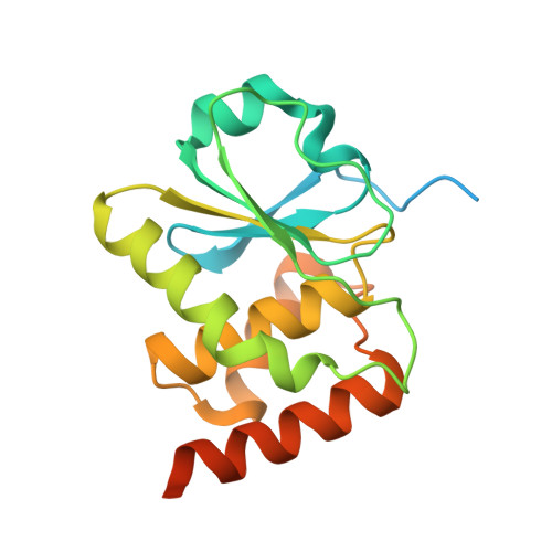
    Dual-specificity phosphatases (DUSPs) constitute a subfamily of protein tyrosine phosphatases, and are intimately involved in the regulation of diverse parameters of cellular signaling and essential biological processes. DUSP28 is one of the DUSP subfamily members that is known to be implicated in the progression of hepatocellular and pancreatic cancers, and its biological functions and enzymatic characteristics are mostly unknown. Herein, we present the crystal structure of human DUSP28 determined to 2.1 Å resolution. DUSP28 adopts a typical DUSP fold, which is composed of a central β-sheet covered by α-helices on both sides and contains a well-ordered activation loop, as do other enzymatically active DUSP proteins. The catalytic pocket of DUSP28, however, appears hardly accessible to a substrate because of the presence of nonconserved bulky residues in the protein tyrosine phosphatase signature motif. Accordingly, DUSP28 showed an atypically low phosphatase activity in the biochemical assay, which was remarkably improved by mutations of two nonconserved residues in the activation loop. Overall, this work reports the structural and biochemical basis for understanding a putative oncological therapeutic target, DUSP28, and also provides a unique mechanism for the regulation of enzymatic activity in the DUSP subfamily proteins.


  • Organizational Affiliation

    Disease Target Structure Research Center, Korea Research Institute of Bioscience and Biotechnology, Daejeon, Republic of Korea.


Macromolecules
Find similar proteins by:  (by identity cutoff)  |  3D Structure
Entity ID: 1
MoleculeChains Sequence LengthOrganismDetailsImage
Dual specificity phosphatase 28
A, B
197Homo sapiensMutation(s): 2 
Gene Names: DUSP28
EC: 3.1.3.16 (PDB Primary Data), 3.1.3.48 (PDB Primary Data)
UniProt & NIH Common Fund Data Resources
Find proteins for Q4G0W2 (Homo sapiens)
Explore Q4G0W2 
Go to UniProtKB:  Q4G0W2
PHAROS:  Q4G0W2
GTEx:  ENSG00000188542 
Entity Groups  
Sequence Clusters30% Identity50% Identity70% Identity90% Identity95% Identity100% Identity
UniProt GroupQ4G0W2
Sequence Annotations
Expand
  • Reference Sequence
Experimental Data & Validation

Experimental Data

  • Method: X-RAY DIFFRACTION
  • Resolution: 2.10 Å
  • R-Value Free: 0.269 
  • R-Value Work: 0.219 
  • R-Value Observed: 0.224 
  • Space Group: P 31 2 1
Unit Cell:
Length ( Å )Angle ( ˚ )
a = 78.952α = 90
b = 78.952β = 90
c = 90.258γ = 120
Software Package:
Software NamePurpose
PHENIXrefinement
HKL-2000data reduction
HKL-2000data scaling
PHENIXphasing

Structure Validation

View Full Validation Report


Entry History & Funding Information

Deposition Data


Funding OrganizationLocationGrant Number
National Research Foundation of KoreaKorea, Republic Of2015M3A9B5030308
National Research Foundation of KoreaKorea, Republic Of2015M3A9B5029741
Korea Research Institute of Bioscience and BiotechnologyKorea, Republic OfResearch Initiative Program

Revision History  (Full details and data files)

  • Version 1.0: 2017-11-22
    Type: Initial release
  • Version 1.1: 2023-11-22
    Changes: Data collection, Database references, Refinement description