


Crystal structure of GH10 xylanase XYL10C from Bispora. sp MEY-1
You, S., Chen, C., Tu, T.To be published.
Experimental Data Snapshot
Entity ID: 1 | |||||
|---|---|---|---|---|---|
| Molecule | Chains | Sequence Length | Organism | Details | Image |
| Beta-xylanase | 335 | Bispora sp. MEY-1 | Mutation(s): 0 Gene Names: xyl10C EC: 3.2.1.8 | 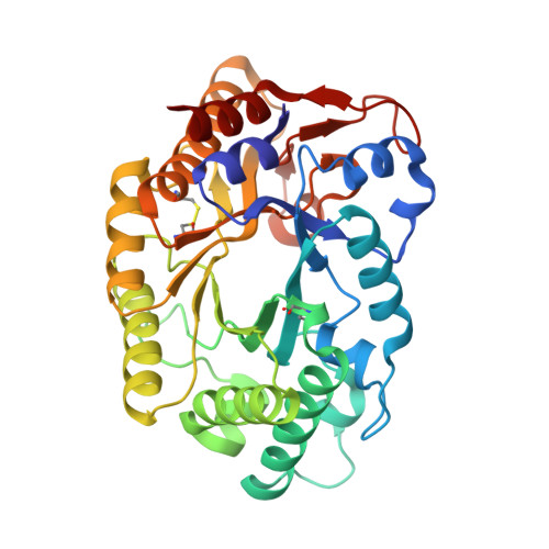 | |
UniProt | |||||
Find proteins for D0QF43 (Bispora sp. MEY-1) Explore D0QF43 Go to UniProtKB: D0QF43 | |||||
Entity Groups | |||||
| Sequence Clusters | 30% Identity50% Identity70% Identity90% Identity95% Identity100% Identity | ||||
| UniProt Group | D0QF43 | ||||
Sequence AnnotationsExpand | |||||
| |||||
| Ligands 1 Unique | |||||
|---|---|---|---|---|---|
| ID | Chains | Name / Formula / InChI Key | 2D Diagram | 3D Interactions | |
| NAG Query on NAG | C [auth A], D [auth A], E [auth A], F [auth B], G [auth B] | 2-acetamido-2-deoxy-beta-D-glucopyranose C8 H15 N O6 OVRNDRQMDRJTHS-FMDGEEDCSA-N |  | ||
| Length ( Å ) | Angle ( ˚ ) |
|---|---|
| a = 135.457 | α = 90 |
| b = 83.262 | β = 94.49 |
| c = 65.279 | γ = 90 |
| Software Name | Purpose |
|---|---|
| PHENIX | refinement |
| xia2 | data reduction |
| xia2 | data scaling |
| PHASER | phasing |
| Funding Organization | Location | Grant Number |
|---|---|---|
| the National Natural Science Foundation of China | China | 31472127 |
RCSB PDB (citation) is hosted by
RCSB PDB is a member of the

