Structural characterization of ribT from Bacillus subtilis reveals it as a GCN5-related N-acetyltransferase.
Srivastava, R., Kaur, A., Sharma, C., Karthikeyan, S.(2018) J Struct Biol 202: 70-81
- PubMed: 29241954
- DOI: https://doi.org/10.1016/j.jsb.2017.12.006
- Primary Citation of Related Structures:
5XXR, 5XXS - PubMed Abstract:
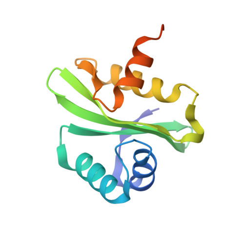
In bacteria, biosynthesis of riboflavin occurs through a series of enzymatic steps starting with one molecule of GTP and two molecules of ribulose-5-phosphate. In Bacillus subtilis (B. subtilis) the genes (ribD/G, ribE, ribA, ribH and ribT) which are involved in riboflavin biosynthesis are organized in an operon referred as rib operon. All the genes of rib operon are characterized functionally except for ribT. The ribT gene with unknown function is found at the distal terminal of rib operon and annotated as a putative N-acetyltransferase. Here, we report the crystal structure of ribT from B. subtilis (bribT) complexed with coenzyme A (CoA) at 2.1 Å resolution determined by single wavelength anomalous dispersion method. Our structural study reveals that bribT is a member of GCN5-related N-acetyltransferase (GNAT) superfamily and contains all the four conserved structural motifs that have been in other members of GNAT superfamily. The members of GNAT family transfers the acetyl group from acetyl coenzyme A (AcCoA) to a variety of substrates. Moreover, the structural analysis reveals that the residues Glu-67 and Ser-107 are suitably positioned to act as a catalytic base and catalytic acid respectively suggesting that the catalysis by bribT may follow a direct transfer mechanism. Surprisingly, the mutation of a non-conserved amino acid residue Cys-112 to alanine or serine affected the binding of AcCoA to bribT, indicating a possible role of Cys-112 in the catalysis.
Organizational Affiliation:
CSIR-Institute of Microbial Technology, Council of Scientific and Industrial Research (CSIR), Sector 39-A, Chandigarh 160 036, India.