5XSD

XylFII-LytSN complex mutant - D103A


Experimental Data Snapshot

  • Method: X-RAY DIFFRACTION
  • Resolution: 2.50 Å
  • R-Value Free: 0.253 
  • R-Value Work: 0.229 
  • R-Value Observed: 0.231 

wwPDB Validation   3D Report Full Report


This is version 1.2 of the entry. See complete history


Literature

Molecular mechanism of environmental d-xylose perception by a XylFII-LytS complex in bacteria

Li, J.Wang, C.Yang, G.Sun, Z.Guo, H.Shao, K.Gu, Y.Jiang, W.Zhang, P.

(2017) Proc Natl Acad Sci U S A 114: 8235-8240

  • DOI: https://doi.org/10.1073/pnas.1620183114
  • Primary Citation of Related Structures:  
    5XSD, 5XSJ, 5XSS

  • PubMed Abstract: 

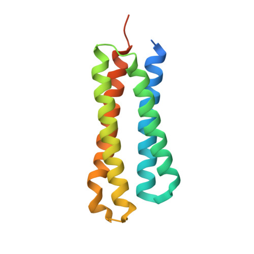
    d-xylose, the main building block of plant biomass, is a pentose sugar that can be used by bacteria as a carbon source for bio-based fuel and chemical production through fermentation. In bacteria, the first step for d-xylose metabolism is signal perception at the membrane. We previously identified a three-component system in Firmicutes bacteria comprising a membrane-associated sensor protein (XylFII), a transmembrane histidine kinase (LytS) for periplasmic d-xylose sensing, and a cytoplasmic response regulator (YesN) that activates the transcription of the target ABC transporter xylFGH genes to promote the uptake of d-xylose. The molecular mechanism underlying signal perception and integration of these processes remains elusive, however. Here we purified the N-terminal periplasmic domain of LytS (LytSN) in a complex with XylFII and determined the conformational structures of the complex in its d-xylose-free and d-xylose-bound forms. LytSN contains a four-helix bundle, and XylFII contains two Rossmann fold-like globular domains with a xylose-binding cleft between them. In the absence of d-xylose, LytSN and XylFII formed a heterodimer. Specific binding of d-xylose to the cleft of XylFII induced a large conformational change that closed the cleft and brought the globular domains closer together. This conformational change led to the formation of an active XylFII-LytSN heterotetramer. Mutations at the d-xylose binding site and the heterotetramer interface diminished heterotetramer formation and impaired the d-xylose-sensing function of XylFII-LytS. Based on these data, we propose a working model of XylFII-LytS that provides a molecular basis for d-xylose utilization and metabolic modification in bacteria.


  • Organizational Affiliation

    National Key Laboratory of Plant Molecular Genetics, Chinese Academy of Sciences Center for Excellence in Molecular Plant Sciences, Institute of Plant Physiology and Ecology, Shanghai Institutes for Biological Sciences, Chinese Academy of Sciences, Shanghai 200032, China.


Macromolecules
Find similar proteins by:  (by identity cutoff)  |  3D Structure
Entity ID: 1
MoleculeChains Sequence LengthOrganismDetailsImage
Periplasmic binding protein/LacI transcriptional regulator
A, B
306Clostridium beijerinckii NCIMB 8052Mutation(s): 1 
Gene Names: Cbei_2377
UniProt
Find proteins for A6LW07 (Clostridium beijerinckii (strain ATCC 51743 / NCIMB 8052))
Explore A6LW07 
Go to UniProtKB:  A6LW07
Entity Groups  
Sequence Clusters30% Identity50% Identity70% Identity90% Identity95% Identity100% Identity
UniProt GroupA6LW07
Sequence Annotations
Expand
  • Reference Sequence
Find similar proteins by:  (by identity cutoff)  |  3D Structure
Entity ID: 2
MoleculeChains Sequence LengthOrganismDetailsImage
Signal transduction histidine kinase, LytSC [auth L]147Clostridium beijerinckii NCIMB 8052Mutation(s): 0 
Gene Names: Cbei_2378
UniProt
Find proteins for A6LW08 (Clostridium beijerinckii (strain ATCC 51743 / NCIMB 8052))
Explore A6LW08 
Go to UniProtKB:  A6LW08
Entity Groups  
Sequence Clusters30% Identity50% Identity70% Identity90% Identity95% Identity100% Identity
UniProt GroupA6LW08
Sequence Annotations
Expand
  • Reference Sequence
Experimental Data & Validation

Experimental Data

  • Method: X-RAY DIFFRACTION
  • Resolution: 2.50 Å
  • R-Value Free: 0.253 
  • R-Value Work: 0.229 
  • R-Value Observed: 0.231 
  • Space Group: P 41 21 2
Unit Cell:
Length ( Å )Angle ( ˚ )
a = 144.958α = 90
b = 144.958β = 90
c = 103.074γ = 90
Software Package:
Software NamePurpose
PHENIXrefinement
HKL-3000data processing
HKL-3000data collection
PHENIXphasing

Structure Validation

View Full Validation Report


Entry History & Funding Information

Deposition Data


Funding OrganizationLocationGrant Number
National Natural Science Foundation of ChinaChina31670755
National Natural Science Foundation of ChinaChina31322016
National Natural Science Foundation of ChinaChina31630003
National Natural Science Foundation of ChinaChina31421061
Chinese Academy of SciencesChinaQYZDB-SSW-SMC006

Revision History  (Full details and data files)

  • Version 1.0: 2017-08-02
    Type: Initial release
  • Version 1.1: 2017-08-09
    Changes: Database references
  • Version 1.2: 2024-03-27
    Changes: Data collection, Database references