


Crystal structure of Carboxyl reductase
Kong, X.D., Xu, J.H., Zhou, J.H.To be published.
Experimental Data Snapshot
wwPDB Validation 3D Report Full Report

Entity ID: 1 | |||||
|---|---|---|---|---|---|
| Molecule | Chains | Sequence Length | Organism | Details | Image |
| Epoxide hydrolase A | 353 | Vigna radiata | Mutation(s): 0 | 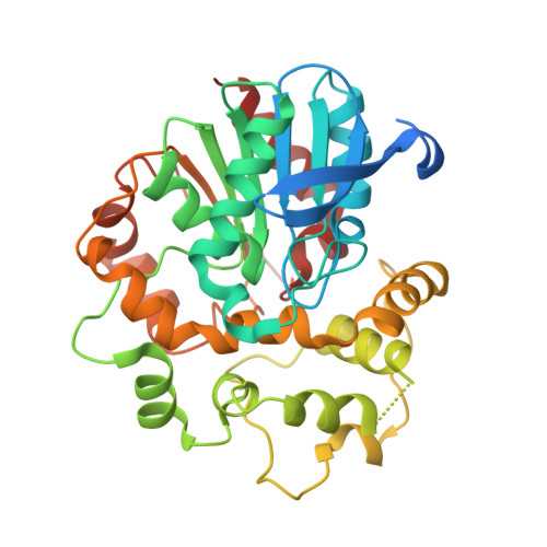 | |
UniProt | |||||
Find proteins for E5L4L1 (Vigna radiata) Explore E5L4L1 Go to UniProtKB: E5L4L1 | |||||
Entity Groups | |||||
| Sequence Clusters | 30% Identity50% Identity70% Identity90% Identity95% Identity100% Identity | ||||
| UniProt Group | E5L4L1 | ||||
Sequence AnnotationsExpand | |||||
| |||||
| Length ( Å ) | Angle ( ˚ ) |
|---|---|
| a = 101.691 | α = 90 |
| b = 51.892 | β = 94.27 |
| c = 124.294 | γ = 90 |
| Software Name | Purpose |
|---|---|
| HKL-2000 | data reduction |
| HKL-2000 | data scaling |
| PHENIX | refinement |
| PHASER | phasing |
| PDB_EXTRACT | data extraction |
| HKL | data reduction |
| HKL | data scaling |
RCSB PDB (citation) is hosted by
RCSB PDB is a member of the

