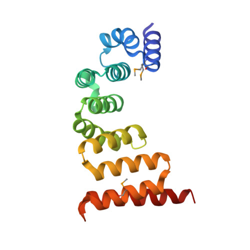
The TPR domain of BepA is required for productive interaction with substrate proteins and the beta-barrel assembly machinery complex.
Daimon, Y., Iwama-Masui, C., Tanaka, Y., Shiota, T., Suzuki, T., Miyazaki, R., Sakurada, H., Lithgow, T., Dohmae, N., Mori, H., Tsukazaki, T., Narita, S.I., Akiyama, Y.(2017) Mol Microbiol 106: 760-776
- PubMed: 28960545
- DOI: https://doi.org/10.1111/mmi.13844
- Primary Citation of Related Structures:
5XI8 - PubMed Abstract:
BepA (formerly YfgC) is an Escherichia coli periplasmic protein consisting of an N-terminal protease domain and a C-terminal tetratricopeptide repeat (TPR) domain. We have previously shown that BepA is a dual functional protein with chaperone-like and proteolytic activities involved in membrane assembly and proteolytic quality control of LptD, a major component of the outer membrane lipopolysaccharide translocon. Intriguingly, BepA can associate with the BAM complex: the β-barrel assembly machinery (BAM) driving integration of β-barrel proteins into the outer membrane. However, the molecular mechanism of BepA function and its association with the BAM complex remains unclear. Here, we determined the crystal structure of the BepA TPR domain, which revealed the presence of two subdomains formed by four TPR motifs. Systematic site-directed in vivo photo-cross-linking was used to map the protein-protein interactions mediated by the BepA TPR domain, showing that this domain interacts both with a substrate and with the BAM complex. Mutational analysis indicated that these interactions are important for the BepA functions. These results suggest that the TPR domain plays critical roles in BepA functions through interactions both with substrates and with the BAM complex. Our findings provide insights into the mechanism of biogenesis and quality control of the outer membrane.
Organizational Affiliation:
Institute for Frontier Life and Medical Sciences, Kyoto University, Kyoto 606-8507, Japan.


