5XGG

Crystal Structure C-terminal SH3 domain of Myosin IB from Entamoeba histolytica


Experimental Data Snapshot

  • Method: X-RAY DIFFRACTION
  • Resolution: 1.72 Å
  • R-Value Free: 0.219 
  • R-Value Work: 0.185 
  • R-Value Observed: 0.186 

wwPDB Validation   3D Report Full Report


This is version 1.1 of the entry. See complete history


Literature

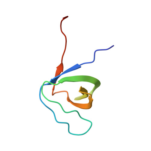
Crystal structure of the PEG-bound SH3 domain of myosin IB from Entamoeba histolytica reveals its mode of ligand recognition

Gautam, G.Rehman, S.A.A.Pandey, P.Gourinath, S.

(2017) Acta Crystallogr D Struct Biol 73: 672-682

  • DOI: https://doi.org/10.1107/S2059798317009639
  • Primary Citation of Related Structures:  
    5XG9, 5XGG

  • PubMed Abstract: 

    The versatility in the recognition of various interacting proteins by the SH3 domain drives a variety of cellular functions. Here, the crystal structure of the C-terminal SH3 domain of myosin IB from Entamoeba histolytica (EhMySH3) is reported at a resolution of 1.7 Å in native and PEG-bound states. Comparisons with other structures indicated that the PEG molecules occupy protein-protein interaction pockets similar to those occupied by the peptides in other peptide-bound SH3-domain structures. Also, analysis of the PEG-bound EhMySH3 structure led to the recognition of two additional pockets, apart from the conventional polyproline and specificity pockets, that are important for ligand interaction. Molecular-docking studies combined with various comparisons revealed structural similarity between EhMySH3 and the SH3 domain of β-Pix, and this similarity led to the prediction that EhMySH3 preferentially binds targets containing type II-like PXXP motifs. These studies expand the understanding of the EhMySH3 domain and provide extensive structural knowledge, which is expected to help in predicting the interacting partners which function together with myosin IB during phagocytosis in E. histolytica infections.


  • Organizational Affiliation

    School of Life Sciences, Jawaharlal Nehru University, New Delhi, Delhi 110 067, India.


Macromolecules
Find similar proteins by:  (by identity cutoff)  |  3D Structure
Entity ID: 1
MoleculeChains Sequence LengthOrganismDetailsImage
Unconventional myosin IB
A, B, C, D, E
A, B, C, D, E, F
66Entamoeba histolyticaMutation(s): 0 
Gene Names: CL6EHI_110810EHI_110810
UniProt
Find proteins for C4LUC7 (Entamoeba histolytica (strain ATCC 30459 / HM-1:IMSS / ABRM))
Explore C4LUC7 
Go to UniProtKB:  C4LUC7
Entity Groups  
Sequence Clusters30% Identity50% Identity70% Identity90% Identity95% Identity100% Identity
UniProt GroupC4LUC7
Sequence Annotations
Expand
  • Reference Sequence
Experimental Data & Validation

Experimental Data

  • Method: X-RAY DIFFRACTION
  • Resolution: 1.72 Å
  • R-Value Free: 0.219 
  • R-Value Work: 0.185 
  • R-Value Observed: 0.186 
  • Space Group: P 1 21 1
Unit Cell:
Length ( Å )Angle ( ˚ )
a = 52.11α = 90
b = 77.67β = 108.24
c = 61.83γ = 90
Software Package:
Software NamePurpose
REFMACrefinement
iMOSFLMdata reduction
SCALEPACKdata scaling
PHASERmodel building

Structure Validation

View Full Validation Report


Entry History & Funding Information

Deposition Data


Funding OrganizationLocationGrant Number
SERBIndia--

Revision History  (Full details and data files)

  • Version 1.0: 2017-08-16
    Type: Initial release
  • Version 1.1: 2023-11-22
    Changes: Data collection, Database references, Refinement description