5XF9

Crystal structure of NAD+-reducing [NiFe]-hydrogenase in the air-oxidized state


Experimental Data Snapshot

  • Method: X-RAY DIFFRACTION
  • Resolution: 2.58 Å
  • R-Value Free: 0.244 
  • R-Value Work: 0.197 
  • R-Value Observed: 0.200 

wwPDB Validation   3D Report Full Report


Ligand Structure Quality Assessment 


This is version 1.2 of the entry. See complete history


Literature

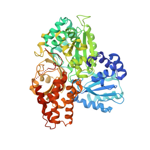
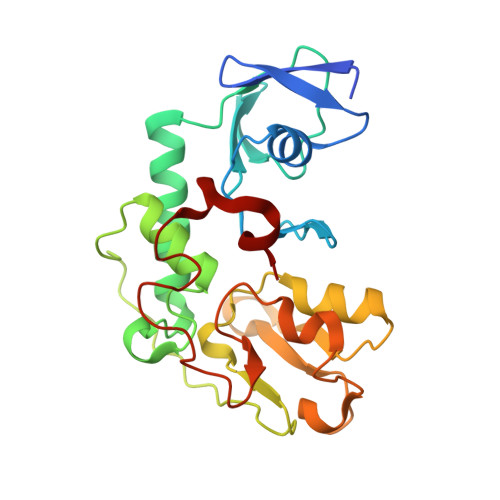
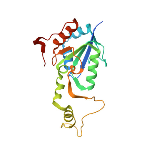
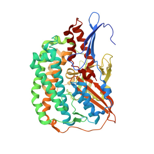
Structural basis of the redox switches in the NAD(+)-reducing soluble [NiFe]-hydrogenase

Shomura, Y.Taketa, M.Nakashima, H.Tai, H.Nakagawa, H.Ikeda, Y.Ishii, M.Igarashi, Y.Nishihara, H.Yoon, K.S.Ogo, S.Hirota, S.Higuchi, Y.

(2017) Science 357: 928-932

  • DOI: https://doi.org/10.1126/science.aan4497
  • Primary Citation of Related Structures:  
    5XF9, 5XFA

  • PubMed Abstract: 

    NAD + (oxidized form of NAD:nicotinamide adenine dinucleotide)-reducing soluble [NiFe]-hydrogenase (SH) is phylogenetically related to NADH (reduced form of NAD + ):quinone oxidoreductase (complex I), but the geometrical arrangements of the subunits and Fe-S clusters are unclear. Here, we describe the crystal structures of SH in the oxidized and reduced states. The cluster arrangement is similar to that of complex I, but the subunits orientation is not, which supports the hypothesis that subunits evolved as prebuilt modules. The oxidized active site includes a six-coordinate Ni, which is unprecedented for hydrogenases, whose coordination geometry would prevent O 2 from approaching. In the reduced state showing the normal active site structure without a physiological electron acceptor, the flavin mononucleotide cofactor is dissociated, which may be caused by the oxidation state change of nearby Fe-S clusters and may suppress production of reactive oxygen species.


  • Organizational Affiliation

    Institute of Quantum Beam Science, Graduate School of Science and Engineering, Ibaraki University, 4-12-1 Nakanarusawa, Hitachi, Ibaraki 316-8511, Japan. hig@sci.u-hyogo.ac.jp yasuhito.shomura.s@vc.ibaraki.ac.jp.


Macromolecules
Find similar proteins by:  (by identity cutoff)  |  3D Structure
Entity ID: 1
MoleculeChains Sequence LengthOrganismDetailsImage
NAD-reducing hydrogenase
A, E
591Hydrogenophilus thermoluteolusMutation(s): 0 
UniProt
Find proteins for A0A077L6X8 (Hydrogenophilus thermoluteolus)
Explore A0A077L6X8 
Go to UniProtKB:  A0A077L6X8
Entity Groups  
Sequence Clusters30% Identity50% Identity70% Identity90% Identity95% Identity100% Identity
UniProt GroupA0A077L6X8
Sequence Annotations
Expand
  • Reference Sequence
Find similar proteins by:  (by identity cutoff)  |  3D Structure
Entity ID: 2
MoleculeChains Sequence LengthOrganismDetailsImage
NAD-reducing hydrogenase
B, F
242Hydrogenophilus thermoluteolusMutation(s): 0 
UniProt
Find proteins for A0A077L885 (Hydrogenophilus thermoluteolus)
Explore A0A077L885 
Go to UniProtKB:  A0A077L885
Entity Groups  
Sequence Clusters30% Identity50% Identity70% Identity90% Identity95% Identity100% Identity
UniProt GroupA0A077L885
Sequence Annotations
Expand
  • Reference Sequence
Find similar proteins by:  (by identity cutoff)  |  3D Structure
Entity ID: 3
MoleculeChains Sequence LengthOrganismDetailsImage
NAD-reducing hydrogenase
C, G
189Hydrogenophilus thermoluteolusMutation(s): 0 
UniProt
Find proteins for A0A077L7R5 (Hydrogenophilus thermoluteolus)
Explore A0A077L7R5 
Go to UniProtKB:  A0A077L7R5
Entity Groups  
Sequence Clusters30% Identity50% Identity70% Identity90% Identity95% Identity100% Identity
UniProt GroupA0A077L7R5
Sequence Annotations
Expand
  • Reference Sequence
Find similar proteins by:  (by identity cutoff)  |  3D Structure
Entity ID: 4
MoleculeChains Sequence LengthOrganismDetailsImage
NAD-reducing hydrogenase
D, H
468Hydrogenophilus thermoluteolusMutation(s): 0 
UniProt
Find proteins for A0A077LAI5 (Hydrogenophilus thermoluteolus)
Explore A0A077LAI5 
Go to UniProtKB:  A0A077LAI5
Entity Groups  
Sequence Clusters30% Identity50% Identity70% Identity90% Identity95% Identity100% Identity
UniProt GroupA0A077LAI5
Sequence Annotations
Expand
  • Reference Sequence
Small Molecules
Ligands 6 Unique
IDChains Name / Formula / InChI Key2D Diagram3D Interactions
FMN
Query on FMN

Download Ideal Coordinates CCD File 
I [auth A],
R [auth E]
FLAVIN MONONUCLEOTIDE
C17 H21 N4 O9 P
FVTCRASFADXXNN-SCRDCRAPSA-N
SF4
Query on SF4

Download Ideal Coordinates CCD File 
J [auth A]
K [auth B]
L [auth B]
N [auth C]
S [auth E]
J [auth A],
K [auth B],
L [auth B],
N [auth C],
S [auth E],
T [auth F],
U [auth F],
W [auth G]
IRON/SULFUR CLUSTER
Fe4 S4
LJBDFODJNLIPKO-UHFFFAOYSA-N
FES
Query on FES

Download Ideal Coordinates CCD File 
M [auth B],
V [auth F]
FE2/S2 (INORGANIC) CLUSTER
Fe2 S2
NIXDOXVAJZFRNF-UHFFFAOYSA-N
FCO
Query on FCO

Download Ideal Coordinates CCD File 
O [auth D],
X [auth H]
CARBONMONOXIDE-(DICYANO) IRON
C3 Fe N2 O
VBQUCMTXYFMTTE-UHFFFAOYSA-N
3NI
Query on 3NI

Download Ideal Coordinates CCD File 
P [auth D],
Y [auth H]
NICKEL (III) ION
Ni
JDRCAGKFDGHRNQ-UHFFFAOYSA-N
MG
Query on MG

Download Ideal Coordinates CCD File 
Q [auth D],
Z [auth H]
MAGNESIUM ION
Mg
JLVVSXFLKOJNIY-UHFFFAOYSA-N
Experimental Data & Validation

Experimental Data

  • Method: X-RAY DIFFRACTION
  • Resolution: 2.58 Å
  • R-Value Free: 0.244 
  • R-Value Work: 0.197 
  • R-Value Observed: 0.200 
  • Space Group: C 1 2 1
Unit Cell:
Length ( Å )Angle ( ˚ )
a = 131.59α = 90
b = 190.09β = 109.44
c = 124.58γ = 90
Software Package:
Software NamePurpose
REFMACrefinement
XDSdata reduction
SCALAdata scaling
PHENIXphasing

Structure Validation

View Full Validation Report


Ligand Structure Quality Assessment 


Entry History & Funding Information

Deposition Data


Funding OrganizationLocationGrant Number
JST CRESTJapanJPMJCR12M4

Revision History  (Full details and data files)

  • Version 1.0: 2017-08-23
    Type: Initial release
  • Version 1.1: 2017-09-13
    Changes: Database references
  • Version 1.2: 2017-09-20
    Changes: Structure summary