5XDM

Structure of the C-terminal domain of E. coli MinC at 3.0 angstrom resolution


Experimental Data Snapshot

  • Method: X-RAY DIFFRACTION
  • Resolution: 3.00 Å
  • R-Value Free: 0.275 
  • R-Value Work: 0.229 
  • R-Value Observed: 0.233 

wwPDB Validation   3D Report Full Report


This is version 1.3 of the entry. See complete history


Literature

Characterization of C-terminal structure of MinC and its implication in evolution of bacterial cell division

Yang, S.Shen, Q.Wang, S.Song, C.Lei, Z.Han, S.Zhang, X.Zheng, J.Jia, Z.

(2017) Sci Rep 7: 7627-7627

  • DOI: https://doi.org/10.1038/s41598-017-08213-5
  • Primary Citation of Related Structures:  
    5XDM

  • PubMed Abstract: 

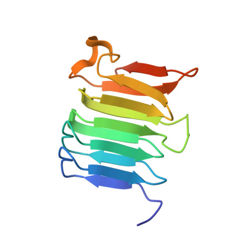
    Proper cell division at the mid-site of Gram-negative bacteria reflects stringent regulation by the min system (MinC, MinD and MinE). Herein we report crystal structure of the C-terminal domain of MinC from Escherichia coli (EcMinC CTD ). The MinC CTD beta helical domain is engaged in a tight homodimer, similar to Thermotoga maritima MinC CTD (TmMinC CTD ). However, both EcMinC CTD and TmMinC CTD lack an α-helix (helix3) at their C-terminal tail, in comparison to Aquifex aerolicu MinC CTD (AaMinC CTD ) which forms an extra interaction interface with MinD. To understand the role of this extra binding element in MinC/MinD interactions, we fused this helix (Aahelix3) to the C-terminus of EcMinC and examined its effect on cell morphology and cell growth. Our results revealed that Aahelix3 impaired normal cell division in vivo. Furthermore, results of a co-pelleting assay and binding free energy calculation suggested that Aahelix3 plays an essential role in AaMinCD complex formation, under the circumstance of lacking MinE in A. aerolicu. Combining these results with sequence analysis of MinC and MinD in different organisms, we propose an evolutionary relationship to rationalize different mechanisms in cell division positioning in various organisms.


  • Organizational Affiliation

    College of Chemistry, Beijing Normal University, Beijing, 100875, China.


Macromolecules
Find similar proteins by:  (by identity cutoff)  |  3D Structure
Entity ID: 1
MoleculeChains Sequence LengthOrganismDetailsImage
Septum site-determining protein MinC
A, B
119Escherichia coli K-12Mutation(s): 0 
Gene Names: minCb1176JW1165
UniProt
Find proteins for P18196 (Escherichia coli (strain K12))
Explore P18196 
Go to UniProtKB:  P18196
Entity Groups  
Sequence Clusters30% Identity50% Identity70% Identity90% Identity95% Identity100% Identity
UniProt GroupP18196
Sequence Annotations
Expand
  • Reference Sequence
Experimental Data & Validation

Experimental Data

  • Method: X-RAY DIFFRACTION
  • Resolution: 3.00 Å
  • R-Value Free: 0.275 
  • R-Value Work: 0.229 
  • R-Value Observed: 0.233 
  • Space Group: P 43 2 2
Unit Cell:
Length ( Å )Angle ( ˚ )
a = 65.061α = 90
b = 65.061β = 90
c = 115.926γ = 90
Software Package:
Software NamePurpose
PHENIXrefinement
HKL-3000data reduction
HKL-3000data scaling
PHENIXphasing

Structure Validation

View Full Validation Report


Entry History & Funding Information

Deposition Data


Funding OrganizationLocationGrant Number
National Natural Science Foundation of ChinaChinaNo. 21273023

Revision History  (Full details and data files)

  • Version 1.0: 2018-02-07
    Type: Initial release
  • Version 1.1: 2018-04-11
    Changes: Data collection, Database references, Refinement description, Source and taxonomy, Structure summary
  • Version 1.2: 2018-04-18
    Changes: Data collection, Source and taxonomy
  • Version 1.3: 2023-11-22
    Changes: Data collection, Database references, Refinement description