5XCZ

Structure of the cellobiohydrolase Cel6A from Phanerochaete chrysosporium in complex with cellobiose at 2.1 angstrom


Experimental Data Snapshot

  • Method: X-RAY DIFFRACTION
  • Resolution: 2.10 Å
  • R-Value Free: 0.237 
  • R-Value Work: 0.178 
  • R-Value Observed: 0.181 

wwPDB Validation   3D Report Full Report


This is version 2.0 of the entry. See complete history


Literature

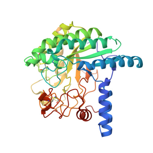
Crystal structure of a family 6 cellobiohydrolase from the basidiomycete Phanerochaete chrysosporium

Tachioka, M.Nakamura, A.Ishida, T.Igarashi, K.Samejima, M.

(2017) Acta Crystallogr F Struct Biol Commun 73: 398-403

  • DOI: https://doi.org/10.1107/S2053230X17008093
  • Primary Citation of Related Structures:  
    5XCY, 5XCZ

  • PubMed Abstract: 

    Cellobiohydrolases belonging to glycoside hydrolase family 6 (CBH II, Cel6A) play key roles in the hydrolysis of crystalline cellulose. CBH II from the white-rot fungus Phanerochaete chrysosporium (PcCel6A) consists of a catalytic domain (CD) and a carbohydrate-binding module connected by a linker peptide, like other known fungal cellobiohydrolases. In the present study, the CD of PcCel6A was crystallized without ligands, and p-nitrophenyl β-D-cellotrioside (pNPG3) was soaked into the crystals. The determined structures of the ligand-free and pNPG3-soaked crystals revealed that binding of cellobiose at substrate subsites +1 and +2 induces a conformational change of the N-terminal and C-terminal loops, switching the tunnel-shaped active site from the open to the closed form.


  • Organizational Affiliation

    Department of Biomaterial Sciences, Graduate School of Agricultural and Life Sciences, The University of Tokyo, 1-1-1 Yayoi, Bunkyo-ku, Tokyo 113-8657, Japan.


Macromolecules
Find similar proteins by:  (by identity cutoff)  |  3D Structure
Entity ID: 1
MoleculeChains Sequence LengthOrganismDetailsImage
Glucanase358Phanerodontia chrysosporiumMutation(s): 0 
Gene Names: cel6A
EC: 3.2.1
UniProt
Find proteins for Q02321 (Phanerodontia chrysosporium)
Explore Q02321 
Go to UniProtKB:  Q02321
Entity Groups  
Sequence Clusters30% Identity50% Identity70% Identity90% Identity95% Identity100% Identity
UniProt GroupQ02321
Sequence Annotations
Expand
  • Reference Sequence
Oligosaccharides

Help

Entity ID: 2
MoleculeChains Length2D Diagram Glycosylation3D Interactions
beta-D-glucopyranose-(1-4)-alpha-D-glucopyranose
B
2N/A
Glycosylation Resources
GlyTouCan:  G86802XL
GlyCosmos:  G86802XL
Small Molecules
Ligands 1 Unique
IDChains Name / Formula / InChI Key2D Diagram3D Interactions
TRS
Query on TRS

Download Ideal Coordinates CCD File 
C [auth A]2-AMINO-2-HYDROXYMETHYL-PROPANE-1,3-DIOL
C4 H12 N O3
LENZDBCJOHFCAS-UHFFFAOYSA-O
Biologically Interesting Molecules (External Reference) 1 Unique
Experimental Data & Validation

Experimental Data

  • Method: X-RAY DIFFRACTION
  • Resolution: 2.10 Å
  • R-Value Free: 0.237 
  • R-Value Work: 0.178 
  • R-Value Observed: 0.181 
  • Space Group: P 21 21 21
Unit Cell:
Length ( Å )Angle ( ˚ )
a = 54.502α = 90
b = 66.951β = 90
c = 85.116γ = 90
Software Package:
Software NamePurpose
PHENIXrefinement
HKL-2000data reduction
HKL-2000data scaling
MOLREPphasing

Structure Validation

View Full Validation Report


Entry History 

Deposition Data

Revision History  (Full details and data files)

  • Version 1.0: 2017-07-26
    Type: Initial release
  • Version 2.0: 2020-07-29
    Type: Remediation
    Reason: Carbohydrate remediation
    Changes: Atomic model, Data collection, Derived calculations, Structure summary