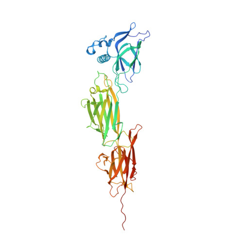
Structures of major pilins in Clostridium perfringens demonstrate dynamic conformational change.
Tamai, E., Katayama, S., Sekiya, H., Nariya, H., Kamitori, S.(2019) Acta Crystallogr D Struct Biol 75: 718-732
- PubMed: 31373571
- DOI: https://doi.org/10.1107/S2059798319009689
- Primary Citation of Related Structures:
5XCB, 5XCC, 6IXY, 6IXZ - PubMed Abstract:
Pili in Gram-positive bacteria are flexible rod proteins associated with the bacterial cell surface, and they play important roles in the initial adhesion to host tissues and colonization. The pilus shaft is formed by the covalent polymerization of major pilins, catalyzed by sortases, a family of cysteine transpeptidases. Here, X-ray structures of the major pilins from Clostridium perfringens strains 13 and SM101 and of sortase from strain SM101 are presented with biochemical analysis to detect the formation of pili in vivo. The major pilin from strain 13 adopts an elongated structure to form noncovalently linked polymeric chains in the crystal, yielding a practical model of the pilus fiber structure. The major pilin from strain SM101 adopts a novel bent structure and associates to form a left-handed twist like an antiparallel double helix in the crystal, which is likely to promote bacterial cell-cell interactions. A modeling study showed that pilin with a bent structure interacts favorably with sortase. The major pilin from strain SM101 was considered to be in an equilibrium state between an elongated and a bent structure through dynamic conformational change, which may be involved in pili-mediated colonization and sortase-mediated polymerization of pili.
Organizational Affiliation:
Department of Infectious Disease, College of Pharmaceutical Science, Matsuyama University, 4-2 Bunkyo-cho, Matsuyama, Ehime 790-8578, Japan.