5XAU

Crystal structure of integrin binding fragment of laminin-511


Experimental Data Snapshot

  • Method: X-RAY DIFFRACTION
  • Resolution: 1.80 Å
  • R-Value Free: 0.237 
  • R-Value Work: 0.202 
  • R-Value Observed: 0.203 

wwPDB Validation   3D Report Full Report


Ligand Structure Quality Assessment 


This is version 2.0 of the entry. See complete history


Literature

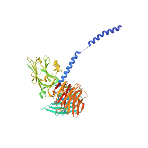
Mechanistic basis for the recognition of laminin-511 by alpha 6 beta 1 integrin.

Takizawa, M.Arimori, T.Taniguchi, Y.Kitago, Y.Yamashita, E.Takagi, J.Sekiguchi, K.

(2017) Sci Adv 3: e1701497-e1701497

  • DOI: https://doi.org/10.1126/sciadv.1701497
  • Primary Citation of Related Structures:  
    5XAU

  • PubMed Abstract: 

    Laminins regulate diverse cellular functions through interaction with integrins. Two regions of laminins-three laminin globular domains of the α chain (LG1-3) and the carboxyl-terminal tail of the γ chain (γ-tail)-are required for integrin binding, but it remains unclear how the γ-tail contributes to the binding. We determined the crystal structure of the integrin binding fragment of laminin-511, showing that the γ-tail extends to the bottom face of LG1-3. Electron microscopic imaging combined with biochemical analyses showed that integrin binds to the bottom face of LG1-3 with the γ1-tail apposed to the metal ion-dependent adhesion site (MIDAS) of integrin β1. These findings are consistent with a model in which the γ-tail coordinates the metal ion in the MIDAS through its Glu residue.


  • Organizational Affiliation

    Division of Matrixome Research and Application, Institute for Protein Research, Osaka University, 3-2 Yamadaoka, Suita, Osaka 565-0871, Japan.


Macromolecules
Find similar proteins by:  (by identity cutoff)  |  3D Structure
Entity ID: 1
MoleculeChains Sequence LengthOrganismDetailsImage
Laminin subunit alpha-5
A, D
674Homo sapiensMutation(s): 1 
Gene Names: LAMA5KIAA0533KIAA1907
UniProt & NIH Common Fund Data Resources
Find proteins for O15230 (Homo sapiens)
Explore O15230 
Go to UniProtKB:  O15230
PHAROS:  O15230
GTEx:  ENSG00000130702 
Entity Groups  
Sequence Clusters30% Identity50% Identity70% Identity90% Identity95% Identity100% Identity
UniProt GroupO15230
Sequence Annotations
Expand
  • Reference Sequence
Find similar proteins by:  (by identity cutoff)  |  3D Structure
Entity ID: 2
MoleculeChains Sequence LengthOrganismDetailsImage
Laminin subunit beta-1
B, E
74Homo sapiensMutation(s): 0 
Gene Names: LAMB1
UniProt & NIH Common Fund Data Resources
Find proteins for P07942 (Homo sapiens)
Explore P07942 
Go to UniProtKB:  P07942
PHAROS:  P07942
GTEx:  ENSG00000091136 
Entity Groups  
Sequence Clusters30% Identity50% Identity70% Identity90% Identity95% Identity100% Identity
UniProt GroupP07942
Sequence Annotations
Expand
  • Reference Sequence
Find similar proteins by:  (by identity cutoff)  |  3D Structure
Entity ID: 3
MoleculeChains Sequence LengthOrganismDetailsImage
Laminin subunit gamma-1
C, F
83Homo sapiensMutation(s): 1 
Gene Names: LAMC1LAMB2
UniProt & NIH Common Fund Data Resources
Find proteins for P11047 (Homo sapiens)
Explore P11047 
Go to UniProtKB:  P11047
PHAROS:  P11047
GTEx:  ENSG00000135862 
Entity Groups  
Sequence Clusters30% Identity50% Identity70% Identity90% Identity95% Identity100% Identity
UniProt GroupP11047
Sequence Annotations
Expand
  • Reference Sequence
Oligosaccharides

Help

Entity ID: 4
MoleculeChains Length2D Diagram Glycosylation3D Interactions
2-acetamido-2-deoxy-beta-D-glucopyranose-(1-4)-2-acetamido-2-deoxy-beta-D-glucopyranose
G
2N-Glycosylation
Glycosylation Resources
GlyTouCan:  G42666HT
GlyCosmos:  G42666HT
GlyGen:  G42666HT
Small Molecules
Ligands 2 Unique
IDChains Name / Formula / InChI Key2D Diagram3D Interactions
NAG
Query on NAG

Download Ideal Coordinates CCD File 
I [auth A]
J [auth A]
K [auth A]
M [auth D]
N [auth D]
I [auth A],
J [auth A],
K [auth A],
M [auth D],
N [auth D],
O [auth D],
P [auth D]
2-acetamido-2-deoxy-beta-D-glucopyranose
C8 H15 N O6
OVRNDRQMDRJTHS-FMDGEEDCSA-N
CA
Query on CA

Download Ideal Coordinates CCD File 
H [auth A],
L [auth D]
CALCIUM ION
Ca
BHPQYMZQTOCNFJ-UHFFFAOYSA-N
Experimental Data & Validation

Experimental Data

  • Method: X-RAY DIFFRACTION
  • Resolution: 1.80 Å
  • R-Value Free: 0.237 
  • R-Value Work: 0.202 
  • R-Value Observed: 0.203 
  • Space Group: C 1 2 1
Unit Cell:
Length ( Å )Angle ( ˚ )
a = 175.031α = 90
b = 121.618β = 127.57
c = 107.554γ = 90
Software Package:
Software NamePurpose
REFMACrefinement
HKL-2000data reduction
HKL-2000data scaling
PHENIXphasing

Structure Validation

View Full Validation Report


Ligand Structure Quality Assessment 


Entry History 

Deposition Data

Revision History  (Full details and data files)

  • Version 1.0: 2017-09-20
    Type: Initial release
  • Version 2.0: 2020-07-29
    Type: Remediation
    Reason: Carbohydrate remediation
    Changes: Atomic model, Data collection, Derived calculations, Structure summary