5X7R

Crystal structure of Paenibacillus sp. 598K alpha-1,6-glucosyltransferase complexed with isomaltohexaose


Experimental Data Snapshot

  • Method: X-RAY DIFFRACTION
  • Resolution: 1.95 Å
  • R-Value Free: 0.208 
  • R-Value Work: 0.178 
  • R-Value Observed: 0.180 

wwPDB Validation   3D Report Full Report


Ligand Structure Quality Assessment 


This is version 2.1 of the entry. See complete history


Literature

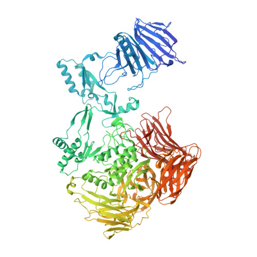
Carbohydrate-binding architecture of the multi-modular alpha-1,6-glucosyltransferase from Paenibacillus sp. 598K, which produces alpha-1,6-glucosyl-alpha-glucosaccharides from starch

Fujimoto, Z.Suzuki, N.Kishine, N.Ichinose, H.Momma, M.Kimura, A.Funane, K.

(2017) Biochem J 474: 2763-2778

  • DOI: https://doi.org/10.1042/BCJ20170152
  • Primary Citation of Related Structures:  
    5X7O, 5X7P, 5X7Q, 5X7R, 5X7S

  • PubMed Abstract: 

    Paenibacillus sp. 598K α-1,6-glucosyltransferase (Ps6TG31A), a member of glycoside hydrolase family 31, catalyzes exo-α-glucohydrolysis and transglucosylation and produces α-1,6-glucosyl-α-glucosaccharides from α-glucan via its disproportionation activity. The crystal structure of Ps6TG31A was determined by an anomalous dispersion method using a terbium derivative. The monomeric Ps6TG31A consisted of one catalytic (β/α) 8 -barrel domain and six small domains, one on the N-terminal and five on the C-terminal side. The structures of the enzyme complexed with maltohexaose, isomaltohexaose, and acarbose demonstrated that the ligands were observed in the catalytic cleft and the sugar-binding sites of four β-domains. The catalytic site was structured by a glucose-binding pocket and an aglycon-binding cleft built by two sidewalls. The bound acarbose was located with its non-reducing end pseudosugar docked in the pocket, and the other moieties along one sidewall serving three subsites for the α-1,4-glucan. The bound isomaltooligosaccharide was found on the opposite sidewall, which provided the space for the acceptor molecule to be positioned for attack of the catalytic intermediate covalent complex during transglucosylation. The N-terminal domain recognized the α-1,4-glucan in a surface-binding mode. Two of the five C-terminal domains belong to the carbohydrate-binding modules family 35 and one to family 61. The sugar complex structures indicated that the first family 35 module preferred α-1,6-glucan, whereas the second family 35 module and family 61 module preferred α-1,4-glucan. Ps6TG31A appears to have enhanced transglucosylation activity facilitated by its carbohydrate-binding modules and substrate-binding cleft that positions the substrate and acceptor sugar for the transglucosylation.


  • Organizational Affiliation

    Advanced Analysis Center, National Agriculture and Food Research Organization (NARO), 2-1-2 Kannondai, Tsukuba, Ibaraki 305-8602, Japan zui@affrc.go.jp funane@affrc.go.jp.


Macromolecules
Find similar proteins by:  (by identity cutoff)  |  3D Structure
Entity ID: 1
MoleculeChains Sequence LengthOrganismDetailsImage
Glycoside hydrolase family 31 alpha-glucosidase
A, B
1,263Paenibacillus sp. 598KMutation(s): 0 
Gene Names: 6gt31a
EC: 2.4.1 (PDB Primary Data), 3.2.1.20 (PDB Primary Data)
UniProt
Find proteins for A0A193PKW5 (Paenibacillus sp. 598K)
Explore A0A193PKW5 
Go to UniProtKB:  A0A193PKW5
Entity Groups  
Sequence Clusters30% Identity50% Identity70% Identity90% Identity95% Identity100% Identity
UniProt GroupA0A193PKW5
Sequence Annotations
Expand
  • Reference Sequence
Oligosaccharides

Help

Entity ID: 2
MoleculeChains Length2D Diagram Glycosylation3D Interactions
alpha-D-glucopyranose-(1-6)-alpha-D-glucopyranose-(1-6)-alpha-D-glucopyranose-(1-6)-alpha-D-glucopyranose-(1-6)-beta-D-glucopyranose
C
5N/A
Glycosylation Resources
GlyTouCan:  G11329BK
GlyCosmos:  G11329BK
Entity ID: 3
MoleculeChains Length2D Diagram Glycosylation3D Interactions
alpha-D-glucopyranose-(1-6)-alpha-D-glucopyranose-(1-6)-alpha-D-glucopyranose
D, F
3N/A
Glycosylation Resources
GlyTouCan:  G02998EX
GlyCosmos:  G02998EX
GlyGen:  G02998EX
Entity ID: 4
MoleculeChains Length2D Diagram Glycosylation3D Interactions
alpha-D-glucopyranose-(1-4)-alpha-D-glucopyranose
E, G
2N/A
Glycosylation Resources
GlyTouCan:  G07411ON
GlyCosmos:  G07411ON
Entity ID: 5
MoleculeChains Length2D Diagram Glycosylation3D Interactions
4,6-dideoxy-4-{[(1S,4R,5S,6S)-4,5,6-trihydroxy-3-(hydroxymethyl)cyclohex-2-en-1-yl]amino}-alpha-D-glucopyranose-(1-4)-alpha-D-glucopyranose
H, I
2N/AN/A
Glycosylation Resources
GlyTouCan:  G61060BJ
GlyCosmos:  G61060BJ
Small Molecules
Ligands 8 Unique
IDChains Name / Formula / InChI Key2D Diagram3D Interactions
MES
Query on MES

Download Ideal Coordinates CCD File 
DA [auth A]
EA [auth A]
FA [auth A]
HA [auth A]
IA [auth A]
DA [auth A],
EA [auth A],
FA [auth A],
HA [auth A],
IA [auth A],
JB [auth B],
KA [auth A],
LA [auth A],
MA [auth A],
OB [auth B],
PB [auth B],
QB [auth B],
TB [auth B],
UB [auth B],
WB [auth B],
YB [auth B]
2-(N-MORPHOLINO)-ETHANESULFONIC ACID
C6 H13 N O4 S
SXGZJKUKBWWHRA-UHFFFAOYSA-N
GLC
Query on GLC

Download Ideal Coordinates CCD File 
N [auth A],
WA [auth B]
alpha-D-glucopyranose
C6 H12 O6
WQZGKKKJIJFFOK-DVKNGEFBSA-N
BGC
Query on BGC

Download Ideal Coordinates CCD File 
M [auth A],
VA [auth B]
beta-D-glucopyranose
C6 H12 O6
WQZGKKKJIJFFOK-VFUOTHLCSA-N
SO4
Query on SO4

Download Ideal Coordinates CCD File 
AA [auth A]
AC [auth B]
BA [auth A]
CA [auth A]
GA [auth A]
AA [auth A],
AC [auth B],
BA [auth A],
CA [auth A],
GA [auth A],
JA [auth A],
KB [auth B],
LB [auth B],
MB [auth B],
NA [auth A],
NB [auth B],
RB [auth B],
SB [auth B],
VB [auth B],
XB [auth B],
ZB [auth B]
SULFATE ION
O4 S
QAOWNCQODCNURD-UHFFFAOYSA-L
EDO
Query on EDO

Download Ideal Coordinates CCD File 
BC [auth B]
CC [auth B]
DC [auth B]
EC [auth B]
OA [auth A]
BC [auth B],
CC [auth B],
DC [auth B],
EC [auth B],
OA [auth A],
PA [auth A],
QA [auth A],
RA [auth A]
1,2-ETHANEDIOL
C2 H6 O2
LYCAIKOWRPUZTN-UHFFFAOYSA-N
NI
Query on NI

Download Ideal Coordinates CCD File 
O [auth A],
P [auth A],
XA [auth B],
YA [auth B]
NICKEL (II) ION
Ni
VEQPNABPJHWNSG-UHFFFAOYSA-N
CA
Query on CA

Download Ideal Coordinates CCD File 
J [auth A]
K [auth A]
L [auth A]
SA [auth B]
TA [auth B]
J [auth A],
K [auth A],
L [auth A],
SA [auth B],
TA [auth B],
UA [auth B]
CALCIUM ION
Ca
BHPQYMZQTOCNFJ-UHFFFAOYSA-N
MG
Query on MG

Download Ideal Coordinates CCD File 
AB [auth B]
BB [auth B]
CB [auth B]
DB [auth B]
EB [auth B]
AB [auth B],
BB [auth B],
CB [auth B],
DB [auth B],
EB [auth B],
FB [auth B],
GB [auth B],
HB [auth B],
IB [auth B],
Q [auth A],
R [auth A],
S [auth A],
T [auth A],
U [auth A],
V [auth A],
W [auth A],
X [auth A],
Y [auth A],
Z [auth A],
ZA [auth B]
MAGNESIUM ION
Mg
JLVVSXFLKOJNIY-UHFFFAOYSA-N
Experimental Data & Validation

Experimental Data

  • Method: X-RAY DIFFRACTION
  • Resolution: 1.95 Å
  • R-Value Free: 0.208 
  • R-Value Work: 0.178 
  • R-Value Observed: 0.180 
  • Space Group: C 2 2 21
Unit Cell:
Length ( Å )Angle ( ˚ )
a = 184.333α = 90
b = 271.26β = 90
c = 133.928γ = 90
Software Package:
Software NamePurpose
REFMACrefinement
HKL-2000data reduction
HKL-2000data scaling
MOLREPphasing

Structure Validation

View Full Validation Report


Ligand Structure Quality Assessment 


Entry History & Funding Information

Deposition Data


Funding OrganizationLocationGrant Number
MAFFJapanA25001

Revision History  (Full details and data files)

  • Version 1.0: 2017-07-26
    Type: Initial release
  • Version 1.1: 2017-08-23
    Changes: Database references
  • Version 1.2: 2017-08-30
    Changes: Data collection
  • Version 2.0: 2020-07-29
    Type: Remediation
    Reason: Carbohydrate remediation
    Changes: Advisory, Atomic model, Data collection, Derived calculations, Non-polymer description, Structure summary
  • Version 2.1: 2023-11-22
    Changes: Data collection, Database references, Derived calculations, Refinement description, Structure summary