5X72

The crystal Structure PDE delta in complex with (rac)-p9


Experimental Data Snapshot

  • Method: X-RAY DIFFRACTION
  • Resolution: 1.95 Å
  • R-Value Free: 0.231 
  • R-Value Work: 0.187 
  • R-Value Observed: 0.190 

wwPDB Validation   3D Report Full Report


Ligand Structure Quality Assessment 


This is version 1.2 of the entry. See complete history


Literature

Structural Biology-Inspired Discovery of Novel KRAS-PDE delta Inhibitors

Jiang, Y.Zhuang, C.Chen, L.Lu, J.Dong, G.Miao, Z.Zhang, W.Li, J.Sheng, C.

(2017) J Med Chem 60: 9400-9406

  • DOI: https://doi.org/10.1021/acs.jmedchem.7b01243
  • Primary Citation of Related Structures:  
    5X72, 5X73, 5X74

  • PubMed Abstract: 

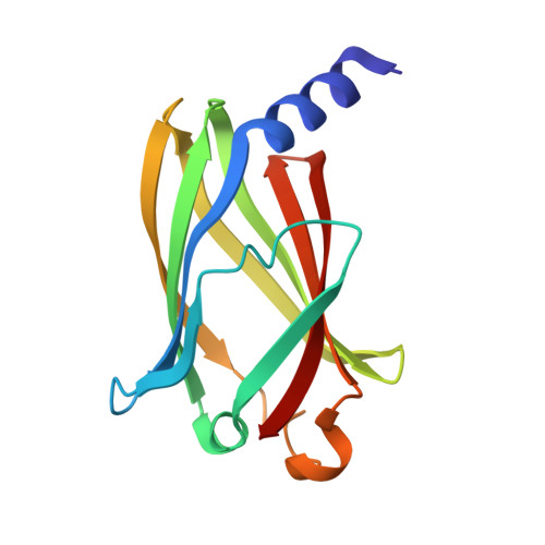
    Structural biology is a powerful tool for investigating the stereospecific interactions between a protein and its ligand. Herein, an unprecedented chiral binding pattern was observed for inhibitors of KRAS-PDEδ interactions. Virtual screening and X-ray crystallography studies revealed that two enantiomers of a racemic inhibitor could bind at different sites. Fragment-based drug design was used to identify highly potent PDEδ inhibitors that can be used as promising lead compounds for target validation and antitumor drug development.


  • Organizational Affiliation

    School of Pharmacy, Second Military Medical University , Shanghai 200433, China.


Macromolecules
Find similar proteins by:  (by identity cutoff)  |  3D Structure
Entity ID: 1
MoleculeChains Sequence LengthOrganismDetailsImage
Retinal rod rhodopsin-sensitive cGMP 3',5'-cyclic phosphodiesterase subunit delta150Homo sapiensMutation(s): 0 
Gene Names: PDE6DPDED
UniProt & NIH Common Fund Data Resources
Find proteins for O43924 (Homo sapiens)
Explore O43924 
Go to UniProtKB:  O43924
PHAROS:  O43924
GTEx:  ENSG00000156973 
Entity Groups  
Sequence Clusters30% Identity50% Identity70% Identity90% Identity95% Identity100% Identity
UniProt GroupO43924
Sequence Annotations
Expand
  • Reference Sequence
Small Molecules
Ligands 2 Unique
IDChains Name / Formula / InChI Key2D Diagram3D Interactions
P69
Query on P69

Download Ideal Coordinates CCD File 
C [auth A](2S)-2-(2-fluorophenyl)-3-phenyl-1,2-dihydroquinazolin-4-one
C20 H15 F N2 O
UMMDFHVNOCVOCD-IBGZPJMESA-N
P59
Query on P59

Download Ideal Coordinates CCD File 
B [auth A](2R)-2-(2-fluorophenyl)-3-phenyl-1,2-dihydroquinazolin-4-one
C20 H15 F N2 O
UMMDFHVNOCVOCD-LJQANCHMSA-N
Binding Affinity Annotations 
IDSourceBinding Affinity
P69 Binding MOAD:  5X72 Kd: 467 (nM) from 1 assay(s)
P59 Binding MOAD:  5X72 Kd: 467 (nM) from 1 assay(s)
Experimental Data & Validation

Experimental Data

  • Method: X-RAY DIFFRACTION
  • Resolution: 1.95 Å
  • R-Value Free: 0.231 
  • R-Value Work: 0.187 
  • R-Value Observed: 0.190 
  • Space Group: P 32 2 1
Unit Cell:
Length ( Å )Angle ( ˚ )
a = 55.568α = 90
b = 55.568β = 90
c = 115.465γ = 120
Software Package:
Software NamePurpose
REFMACrefinement
HKL-2000data reduction
HKL-2000data scaling

Structure Validation

View Full Validation Report


Ligand Structure Quality Assessment 


Entry History & Funding Information

Deposition Data


Funding OrganizationLocationGrant Number
National Basic Research Program of ChinaChina2014CB541800

Revision History  (Full details and data files)

  • Version 1.0: 2017-10-04
    Type: Initial release
  • Version 1.1: 2017-12-06
    Changes: Database references
  • Version 1.2: 2023-11-22
    Changes: Data collection, Database references, Refinement description