5X6J

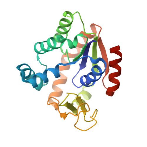
Crystal structure of B. globisporus adenylate kinase variant


Experimental Data Snapshot

  • Method: X-RAY DIFFRACTION
  • Resolution: 2.10 Å
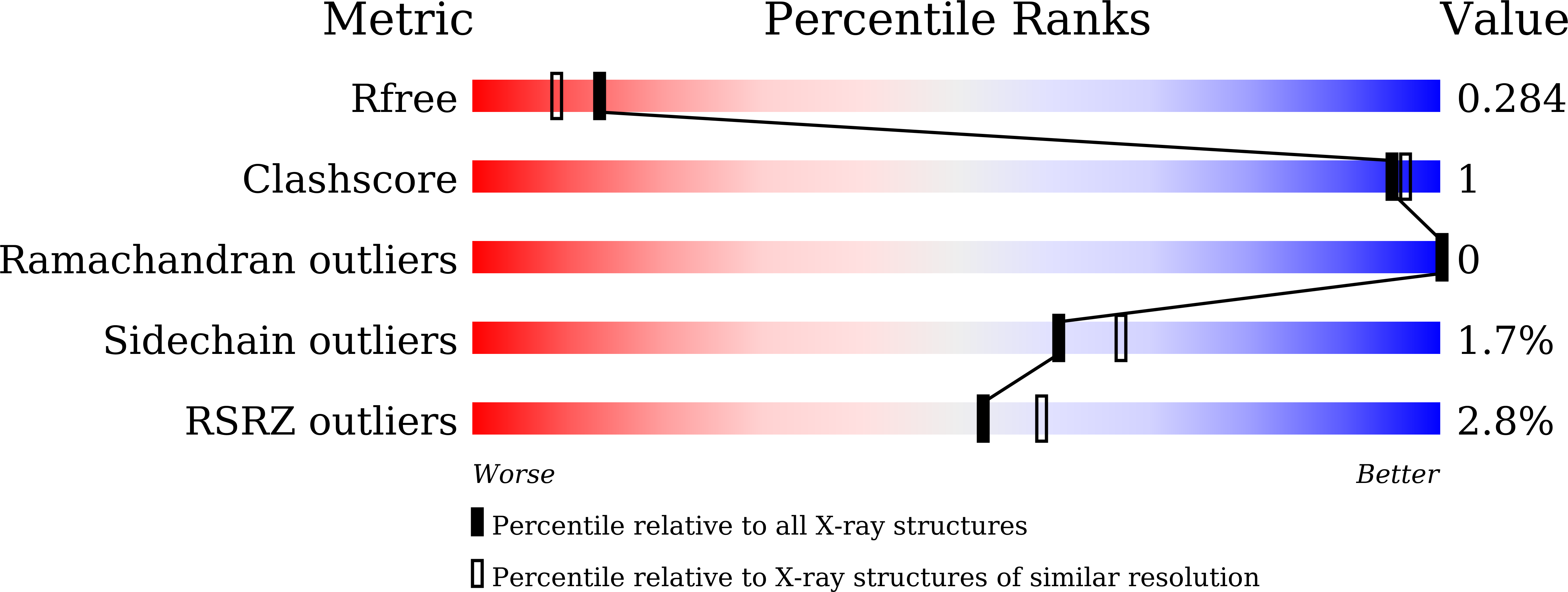
  • R-Value Free: 0.276 
  • R-Value Work: 0.193 
  • R-Value Observed: 0.197 

wwPDB Validation   3D Report Full Report


Ligand Structure Quality Assessment 


This is version 1.2 of the entry. See complete history


Literature

Structural and mutational analyses of psychrophilic and mesophilic adenylate kinases highlight the role of hydrophobic interactions in protein thermal stability.

Moon, S.Kim, J.Koo, J.Bae, E.

(2019) Struct Dyn 6: 024702-024702

  • DOI: https://doi.org/10.1063/1.5089707
  • Primary Citation of Related Structures:  
    5X6I, 5X6J

  • PubMed Abstract: 

    Protein thermal stability is an important field since thermally stable proteins are desirable in many academic and industrial settings. Information on protein thermal stabilization can be obtained by comparing homologous proteins from organisms living at distinct temperatures. Here, we report structural and mutational analyses of adenylate kinases (AKs) from psychrophilic Bacillus globisporus (AKp) and mesophilic Bacillus subtilis (AKm). Sequence and structural comparison showed suboptimal hydrophobic packing around Thr26 in the CORE domain of AKp, which was replaced with an Ile residue in AKm. Mutations that improved hydrophobicity of the Thr residue increased the thermal stability of the psychrophilic AKp, and the largest stabilization was observed for a Thr-to-Ile substitution. Furthermore, a reverse Ile-to-Thr mutation in the mesophilic AKm significantly decreased thermal stability. We determined the crystal structures of mutant AKs to confirm the impact of the residue substitutions on the overall stability. Taken together, our results provide a structural basis for the stability difference between psychrophilic and mesophilic AK homologues and highlight the role of hydrophobic interactions in protein thermal stability.


  • Organizational Affiliation

    Department of Agricultural Biotechnology, Seoul National University, Seoul 08826, South Korea.


Macromolecules
Find similar proteins by:  (by identity cutoff)  |  3D Structure
Entity ID: 1
MoleculeChains Sequence LengthOrganismDetailsImage
Adenylate kinase217Sporosarcina globisporaMutation(s): 1 
Gene Names: adk
EC: 2.7.4.3
UniProt
Find proteins for P84139 (Sporosarcina globispora)
Explore P84139 
Go to UniProtKB:  P84139
Entity Groups  
Sequence Clusters30% Identity50% Identity70% Identity90% Identity95% Identity100% Identity
UniProt GroupP84139
Sequence Annotations
Expand
  • Reference Sequence
Small Molecules
Ligands 2 Unique
IDChains Name / Formula / InChI Key2D Diagram3D Interactions
AP5
Query on AP5

Download Ideal Coordinates CCD File 
C [auth A]BIS(ADENOSINE)-5'-PENTAPHOSPHATE
C20 H29 N10 O22 P5
OIMACDRJUANHTJ-XPWFQUROSA-N
ZN
Query on ZN

Download Ideal Coordinates CCD File 
B [auth A]ZINC ION
Zn
PTFCDOFLOPIGGS-UHFFFAOYSA-N
Experimental Data & Validation

Experimental Data

  • Method: X-RAY DIFFRACTION
  • Resolution: 2.10 Å
  • R-Value Free: 0.276 
  • R-Value Work: 0.193 
  • R-Value Observed: 0.197 
  • Space Group: P 31 2 1
Unit Cell:
Length ( Å )Angle ( ˚ )
a = 65.106α = 90
b = 65.106β = 90
c = 94.565γ = 120
Software Package:
Software NamePurpose
REFMACrefinement
HKL-2000data reduction
HKL-2000data scaling
PHASERphasing

Structure Validation

View Full Validation Report


Ligand Structure Quality Assessment 


Entry History 

Deposition Data

Revision History  (Full details and data files)

  • Version 1.0: 2018-02-28
    Type: Initial release
  • Version 1.1: 2019-09-11
    Changes: Data collection, Database references
  • Version 1.2: 2023-11-22
    Changes: Data collection, Database references, Refinement description