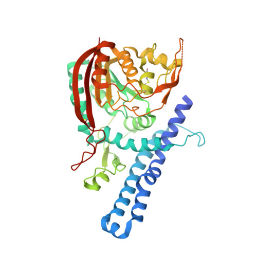
Substrate Recognition Mechanism of the Putative Yeast Carnosine N-methyltransferase
Liu, X., Wu, J., Sun, Y., Xie, W.(2017) ACS Chem Biol 12: 2164-2171
- PubMed: 28654751
- DOI: https://doi.org/10.1021/acschembio.7b00328
- Primary Citation of Related Structures:
5X62 - PubMed Abstract:
Anserine (β-alanyl-N(P i )-methyl-l-histidine) is a natural metabolite present in skeletal muscle and the central nervous system of vertebrates and plays important physiological roles in living organisms. The production of anserine is catalyzed by carnosine N-methyltransferases, which transfer a methyl group to carnosine (β-alanyl-l-histidine). However, the structural basis of the substrate recognition for the enzymes is unknown. We present the crystal structure of the putative carnosine N-methyltransferase from yeast named YNL092W in complex with SAH, solved by the single-wavelength anomalous dispersion (SAD) method. The protein contains a typical Rossmann domain and a characteristic N-terminal helical domain. At the cofactor-binding site, SAH forms an extensive interaction network with the enzyme. The individual contribution of each residue to ligand affinity and enzyme activity was assessed by ITC and methyltransferase assays after mutagenesis of the key residues. Additionally, docking studies and activity assays were conducted in order to identify the binding site for carnosine, and a plausible complex model was proposed. Furthermore, we discovered that two disulfide bridges might be functionally important to the enzyme. By comparison to structure- and sequence-similar methyltransferases, we deduce that the enzyme most likely acts on a protein substrate. Our structural analyses shed light on the catalytic mechanism and substrate recognition by YNL092W.
Organizational Affiliation:
Key Laboratory of Gene Engineering of the Ministry of Education, State Key Laboratory for Biocontrol, School of Life Sciences, The Sun Yat-Sen University , 135 W. Xingang Rd., Guangzhou 510275, People's Republic of China.