5X5I

The X-ray crystal structure of a TetR family transcription regulator RcdA involved in the regulation of biofilm formation in Escherichia coli


Experimental Data Snapshot

  • Method: X-RAY DIFFRACTION
  • Resolution: 2.55 Å
  • R-Value Free: 0.267 
  • R-Value Work: 0.198 
  • R-Value Observed: 0.201 

wwPDB Validation   3D Report Full Report


This is version 1.0 of the entry. See complete history


Literature

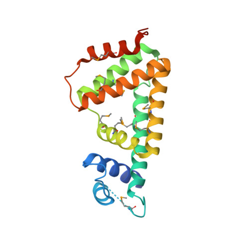
A structural sketch of RcdA, a transcription factor controlling the master regulator of biofilm formation.

Sugino, H.Usui, T.Shimada, T.Nakano, M.Ogasawara, H.Ishihama, A.Hirata, A.

(2017) FEBS Lett 591: 2019-2031

  • DOI: https://doi.org/10.1002/1873-3468.12713
  • Primary Citation of Related Structures:  
    5X5I

  • PubMed Abstract: 

    RcdA is a regulator of curlin subunit gene D, the master regulator of biofilm formation in Escherichia coli. Here, we determined the X-ray structure of RcdA at 2.55 Å resolution. RcdA consists of an N-terminal DNA-binding domain (DBD) containing a helix-turn-helix (HTH) motif and a C-terminal dimerization domain, and forms a homodimer in crystals. A computational docking model of the RcdA-DNA complex allowed prediction of the candidate residues responsible for DNA binding. Our structure-guided mutagenesis, in combination with gel shift assay, atomic force microscopic observation, and reporter assay, indicate that R32 in α2 of the HTH motif plays an essential role in the recognition and binding of target DNA while T46 in α3 influences the mode of oligomerization. These results provide insights into the DNA-binding mode of RcdA.


  • Organizational Affiliation

    Department of Materials Science and Biotechnology, Graduate School of Science and Engineering, Ehime University, Matsuyama, Japan.


Macromolecules
Find similar proteins by:  (by identity cutoff)  |  3D Structure
Entity ID: 1
MoleculeChains Sequence LengthOrganismDetailsImage
HTH-type transcriptional regulator RcdA
A, B, C, D, E
A, B, C, D, E, F, G, H
189Escherichia coli K-12Mutation(s): 0 
Gene Names: rcdAybjKb0846JW5114
UniProt
Find proteins for P75811 (Escherichia coli (strain K12))
Explore P75811 
Go to UniProtKB:  P75811
Entity Groups  
Sequence Clusters30% Identity50% Identity70% Identity90% Identity95% Identity100% Identity
UniProt GroupP75811
Sequence Annotations
Expand
  • Reference Sequence
Small Molecules
Modified Residues  1 Unique
IDChains TypeFormula2D DiagramParent
MSE
Query on MSE
A, B, C, D, E
A, B, C, D, E, F, G, H
L-PEPTIDE LINKINGC5 H11 N O2 SeMET
Experimental Data & Validation

Experimental Data

  • Method: X-RAY DIFFRACTION
  • Resolution: 2.55 Å
  • R-Value Free: 0.267 
  • R-Value Work: 0.198 
  • R-Value Observed: 0.201 
  • Space Group: P 1
Unit Cell:
Length ( Å )Angle ( ˚ )
a = 38.477α = 90
b = 75.967β = 90.01
c = 110.051γ = 89.95
Software Package:
Software NamePurpose
PHENIXrefinement
HKL-2000data reduction
HKL-2000data scaling
PHENIXphasing

Structure Validation

View Full Validation Report


Entry History & Funding Information

Deposition Data


Funding OrganizationLocationGrant Number
Grants-in-Aid for Scientific Research on Innovative Areas JSPS KAKENHIJapan25118516
Scientific Research (C)JSPS KAKENHIJapan15K06975

Revision History  (Full details and data files)

  • Version 1.0: 2017-12-27
    Type: Initial release