5X49

Crystal Structure of Human mitochondrial X-prolyl Aminopeptidase (XPNPEP3)


Experimental Data Snapshot

  • Method: X-RAY DIFFRACTION
  • Resolution: 1.65 Å
  • R-Value Free: 0.177 
  • R-Value Work: 0.154 
  • R-Value Observed: 0.155 

wwPDB Validation   3D Report Full Report


Ligand Structure Quality Assessment 


This is version 2.1 of the entry. See complete history


Literature

Structure of the human aminopeptidase XPNPEP3 and comparison of its in vitro activity with Icp55 orthologs: Insights into diverse cellular processes.

Singh, R.Jamdar, S.N.Goyal, V.D.Kumar, A.Ghosh, B.Makde, R.D.

(2017) J Biol Chem 292: 10035-10047

  • DOI: https://doi.org/10.1074/jbc.M117.783357
  • Primary Citation of Related Structures:  
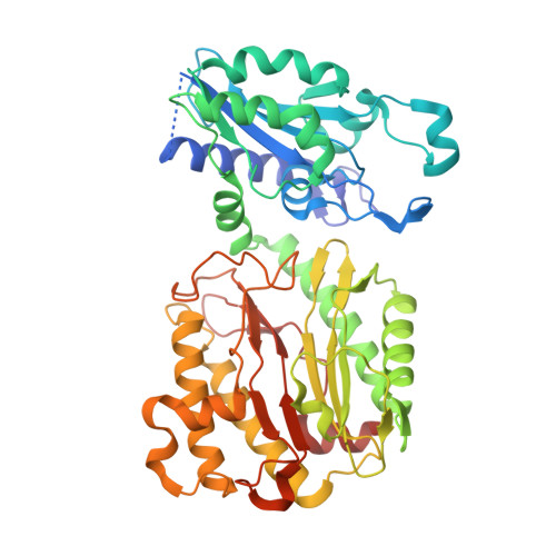
    5X49

  • PubMed Abstract: 

    The human aminopeptidase XPNPEP3 is associated with cystic kidney disease and TNF-TNFR2 cellular signaling. Its yeast and plant homolog Icp55 processes several imported mitochondrial matrix proteins leading to their stabilization. However, the molecular basis for the diverse roles of these enzymes in the cell is unknown. Here, we report the crystal structure of human XPNPEP3 with bound apstatin product at 1.65 Å resolution, and we compare its in vitro substrate specificity with those of fungal Icp55 enzymes. In contrast to the suggestions by earlier in vivo studies of mitochondrial processing, we found that these enzymes are genuine Xaa-Pro aminopeptidases, which hydrolyze peptides with proline at the second position (P1'). The mitochondrial processing activity involving cleavage of peptides lacking P1' proline was also detected in the purified enzymes. A wide proline pocket as well as molecular complementarity and capping at the S1 substrate site of XPNPEP3 provide the necessary structural features for processing the mitochondrial substrates. However, this activity was found to be significantly lower as compared with Xaa-Pro aminopeptidase activity. Because of similar activity profiles of Icp55 and XPNPEP3, we propose that XPNPEP3 plays the same mitochondrial role in humans as Icp55 does in yeast. Both Xaa-Pro aminopeptidase and mitochondrial processing activities of XPNPEP3 have implications toward mitochondrial fitness and cystic kidney disease. Furthermore, the presence of both these activities in Icp55 elucidates the unexplained processing of the mitochondrial cysteine desulfurase Nfs1 in yeast. The enzymatic and structural analyses reported here provide a valuable molecular framework for understanding the diverse cellular roles of XPNPEP3.


  • Organizational Affiliation

    From the High Pressure and Synchrotron Radiation Physics Division and.


Macromolecules
Find similar proteins by:  (by identity cutoff)  |  3D Structure
Entity ID: 1
MoleculeChains Sequence LengthOrganismDetailsImage
Probable Xaa-Pro aminopeptidase 3
A, B
456Homo sapiensMutation(s): 0 
Gene Names: XPNPEP3
EC: 3.4.11.9
UniProt & NIH Common Fund Data Resources
Find proteins for Q9NQH7 (Homo sapiens)
Explore Q9NQH7 
Go to UniProtKB:  Q9NQH7
PHAROS:  Q9NQH7
GTEx:  ENSG00000196236 
Entity Groups  
Sequence Clusters30% Identity50% Identity70% Identity90% Identity95% Identity100% Identity
UniProt GroupQ9NQH7
Sequence Annotations
Expand
  • Reference Sequence
Small Molecules
Ligands 5 Unique
IDChains Name / Formula / InChI Key2D Diagram3D Interactions
12P
Query on 12P

Download Ideal Coordinates CCD File 
E [auth A],
M [auth B]
DODECAETHYLENE GLYCOL
C24 H50 O13
WRZXKWFJEFFURH-UHFFFAOYSA-N
01B
Query on 01B

Download Ideal Coordinates CCD File 
F [auth A],
N [auth B]
(2S,3R)-3-amino-2-hydroxy-4-phenylbutanoic acid
C10 H13 N O3
LDSJMFGYNFIFRK-BDAKNGLRSA-N
DMS
Query on DMS

Download Ideal Coordinates CCD File 
G [auth A]
H [auth A]
I [auth A]
O [auth B]
P [auth B]
G [auth A],
H [auth A],
I [auth A],
O [auth B],
P [auth B],
Q [auth B]
DIMETHYL SULFOXIDE
C2 H6 O S
IAZDPXIOMUYVGZ-UHFFFAOYSA-N
EDO
Query on EDO

Download Ideal Coordinates CCD File 
J [auth A],
R [auth B]
1,2-ETHANEDIOL
C2 H6 O2
LYCAIKOWRPUZTN-UHFFFAOYSA-N
MN
Query on MN

Download Ideal Coordinates CCD File 
C [auth A],
D [auth A],
K [auth B],
L [auth B]
MANGANESE (II) ION
Mn
WAEMQWOKJMHJLA-UHFFFAOYSA-N
Experimental Data & Validation

Experimental Data

  • Method: X-RAY DIFFRACTION
  • Resolution: 1.65 Å
  • R-Value Free: 0.177 
  • R-Value Work: 0.154 
  • R-Value Observed: 0.155 
  • Space Group: P 1 21 1
Unit Cell:
Length ( Å )Angle ( ˚ )
a = 62.897α = 90
b = 135.103β = 99.87
c = 67.205γ = 90
Software Package:
Software NamePurpose
PHENIXrefinement
Cootmodel building
PHENIXmodel building
PHASERphasing
Aimlessdata processing
XDSdata processing
MAR345dtbdata collection

Structure Validation

View Full Validation Report


Ligand Structure Quality Assessment 


Entry History 

Deposition Data

Revision History  (Full details and data files)

  • Version 1.0: 2017-05-17
    Type: Initial release
  • Version 1.1: 2017-12-06
    Changes: Database references
  • Version 2.0: 2023-11-15
    Changes: Atomic model, Data collection, Database references, Derived calculations
  • Version 2.1: 2023-11-22
    Changes: Refinement description