5X3D

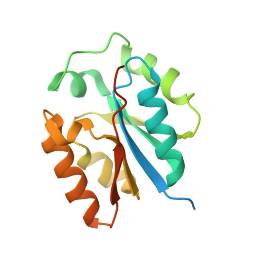
Crystal structure of HEP-CMP-bound form of cytidylyltransferase (CyTase) domain of Fom1 from Streptomyces wedmorensis


Experimental Data Snapshot

  • Method: X-RAY DIFFRACTION
  • Resolution: 1.93 Å
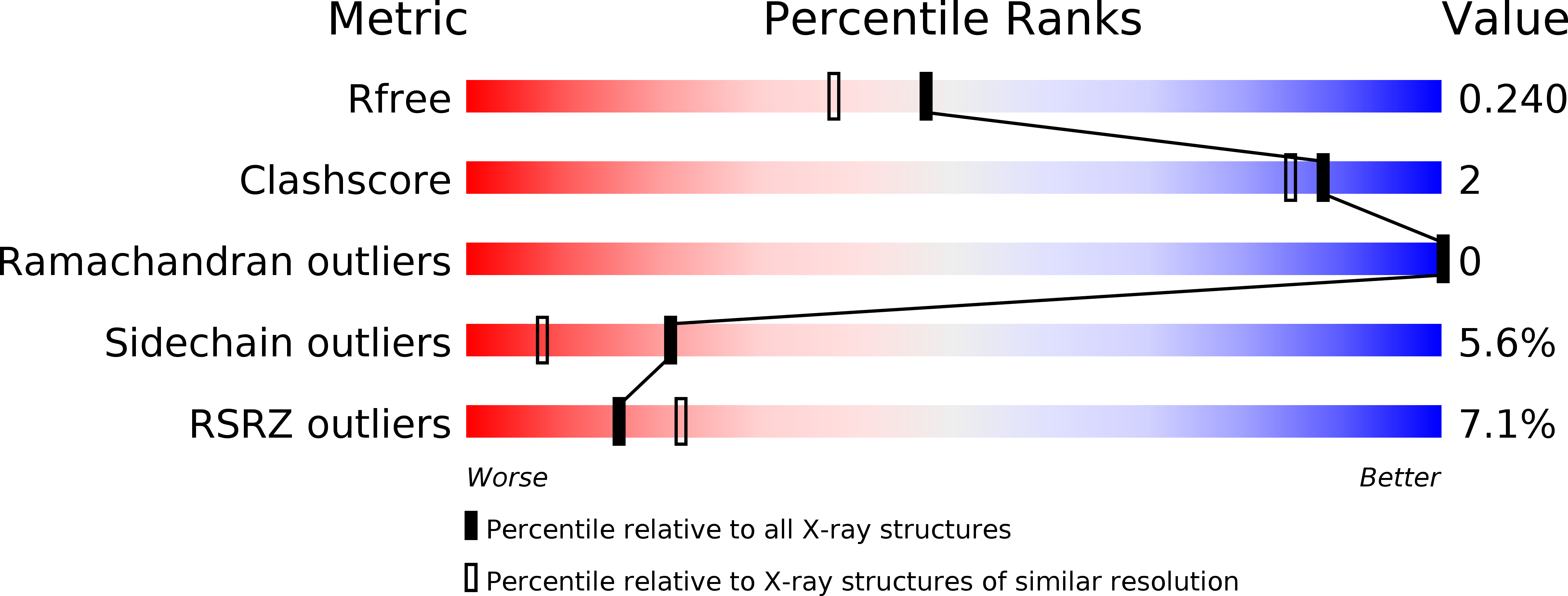
  • R-Value Free: 0.234 
  • R-Value Work: 0.191 
  • R-Value Observed: 0.193 

wwPDB Validation   3D Report Full Report


Ligand Structure Quality Assessment 


This is version 1.1 of the entry. See complete history


Literature

Fosfomycin Biosynthesis via Transient Cytidylylation of 2-Hydroxyethylphosphonate by the Bifunctional Fom1 Enzyme

Cho, S.H.Kim, S.Y.Tomita, T.Shiraishi, T.Park, J.S.Sato, S.Kudo, F.Eguchi, T.Funa, N.Nishiyama, M.Kuzuyama, T.

(2017) ACS Chem Biol 12: 2209-2215

  • DOI: https://doi.org/10.1021/acschembio.7b00419
  • Primary Citation of Related Structures:  
    5X3D

  • PubMed Abstract: 

    Fosfomycin is a wide-spectrum phosphonate antibiotic that is used clinically to treat cystitis, tympanitis, etc. Its biosynthesis starts with the formation of a carbon-phosphorus bond catalyzed by the phosphoenolpyruvate phosphomutase Fom1. We identified an additional cytidylyltransferase (CyTase) domain at the Fom1 N-terminus in addition to the phosphoenolpyruvate phosphomutase domain at the Fom1 C-terminus. Here, we demonstrate that Fom1 is bifunctional and that the Fom1 CyTase domain catalyzes the cytidylylation of the 2-hydroxyethylphosphonate (HEP) intermediate to produce cytidylyl-HEP. On the basis of this new function of Fom1, we propose a revised fosfomycin biosynthetic pathway that involves the transient CMP-conjugated intermediate. The identification of a biosynthetic mechanism via such transient cytidylylation of a biosynthetic intermediate fundamentally advances the understanding of phosphonate biosynthesis in nature. The crystal structure of the cytidylyl-HEP-bound CyTase domain provides a basis for the substrate specificity and reveals unique catalytic elements not found in other members of the CyTase family.


  • Organizational Affiliation

    Biotechnology Research Center, The University of Tokyo , 1-1-1 Yayoi, Bunkyo-ku, Tokyo 113-8657, Japan.


Macromolecules
Find similar proteins by:  (by identity cutoff)  |  3D Structure
Entity ID: 1
MoleculeChains Sequence LengthOrganismDetailsImage
Phosphoenolpyruvate phosphomutase160Streptomyces wedmorensisMutation(s): 0 
Gene Names: Fom1(N)
UniProt
Find proteins for P96074 (Streptomyces wedmorensis)
Explore P96074 
Go to UniProtKB:  P96074
Entity Groups  
Sequence Clusters30% Identity50% Identity70% Identity90% Identity95% Identity100% Identity
UniProt GroupP96074
Sequence Annotations
Expand
  • Reference Sequence
Small Molecules
Ligands 1 Unique
IDChains Name / Formula / InChI Key2D Diagram3D Interactions
7XL
Query on 7XL

Download Ideal Coordinates CCD File 
B [auth A][[(2R,3S,4R,5R)-5-(4-azanyl-2-oxidanylidene-pyrimidin-1-yl)-3,4-bis(oxidanyl)oxolan-2-yl]methoxy-oxidanyl-phosphoryl]oxy-(2-hydroxyethyl)phosphinic acid
C11 H19 N3 O11 P2
ODFOOQGQRDVSPW-PEBGCTIMSA-N
Experimental Data & Validation

Experimental Data

  • Method: X-RAY DIFFRACTION
  • Resolution: 1.93 Å
  • R-Value Free: 0.234 
  • R-Value Work: 0.191 
  • R-Value Observed: 0.193 
  • Space Group: P 65 2 2
Unit Cell:
Length ( Å )Angle ( ˚ )
a = 71.909α = 90
b = 71.909β = 90
c = 109.507γ = 120
Software Package:
Software NamePurpose
REFMACrefinement
HKL-2000data reduction
HKL-2000data scaling
SOLVEphasing

Structure Validation

View Full Validation Report


Ligand Structure Quality Assessment 


Entry History 

Deposition Data

Revision History  (Full details and data files)

  • Version 1.0: 2017-09-13
    Type: Initial release
  • Version 1.1: 2024-03-27
    Changes: Data collection, Database references