


Crystal structure of mouse sulfotransferase SULT7A1 complexed with PAP
Tanaka, S., Kakuta, Y.To be published.
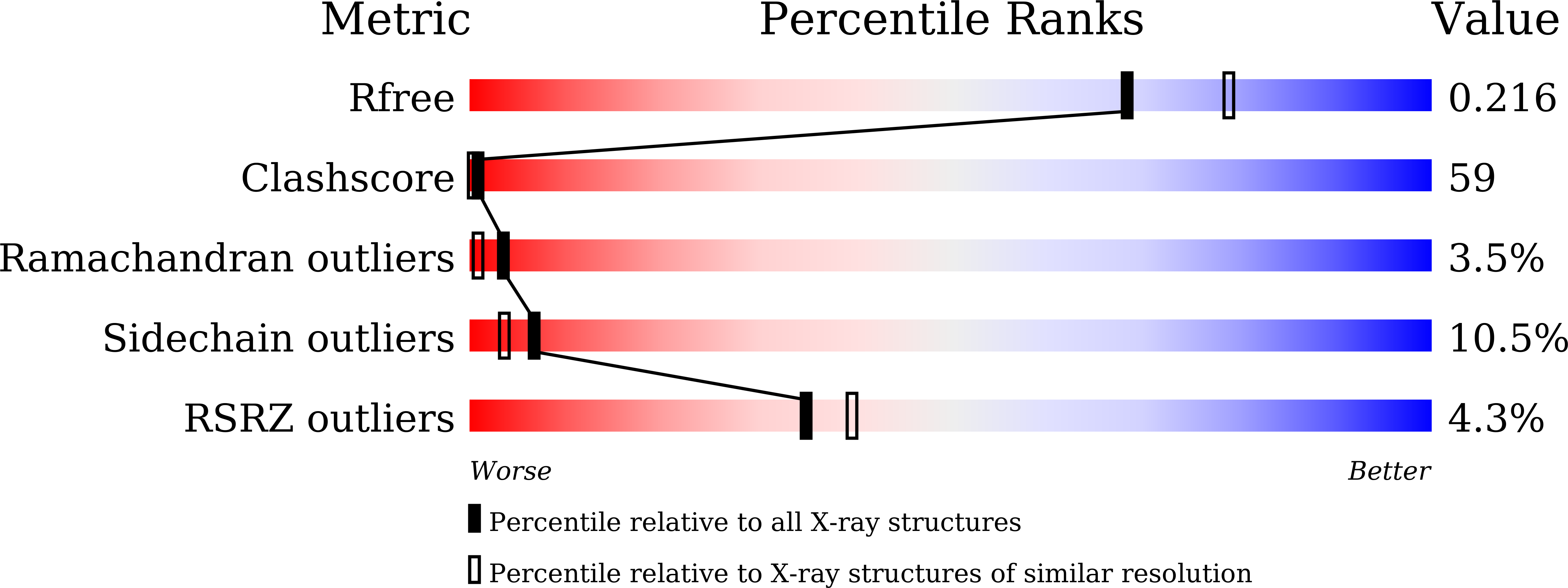
Experimental Data Snapshot
Entity ID: 1 | |||||
|---|---|---|---|---|---|
| Molecule | Chains | Sequence Length | Organism | Details | Image |
| Sulfotransferase | 283 | Mus musculus | Mutation(s): 0 Gene Names: Sult6b2, Gm766 EC: 2.8.2 | 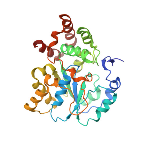 | |
UniProt | |||||
Find proteins for B7ZWN4 (Mus musculus) Explore B7ZWN4 Go to UniProtKB: B7ZWN4 | |||||
Entity Groups | |||||
| Sequence Clusters | 30% Identity50% Identity70% Identity90% Identity95% Identity100% Identity | ||||
| UniProt Group | B7ZWN4 | ||||
Sequence AnnotationsExpand | |||||
| |||||
Entity ID: 2 | |||||
|---|---|---|---|---|---|
| Molecule | Chains | Sequence Length | Organism | Details | Image |
| Sulfotransferase | 281 | Mus musculus | Mutation(s): 0 Gene Names: Sult6b2, Gm766 EC: 2.8.2 | 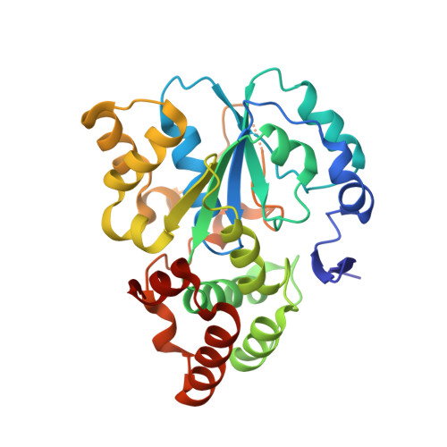 | |
UniProt | |||||
Find proteins for B7ZWN4 (Mus musculus) Explore B7ZWN4 Go to UniProtKB: B7ZWN4 | |||||
Entity Groups | |||||
| Sequence Clusters | 30% Identity50% Identity70% Identity90% Identity95% Identity100% Identity | ||||
| UniProt Group | B7ZWN4 | ||||
Sequence AnnotationsExpand | |||||
| |||||
Entity ID: 3 | |||||
|---|---|---|---|---|---|
| Molecule | Chains | Sequence Length | Organism | Details | Image |
| Sulfotransferase | 282 | Mus musculus | Mutation(s): 0 Gene Names: Sult6b2, Gm766 EC: 2.8.2 | 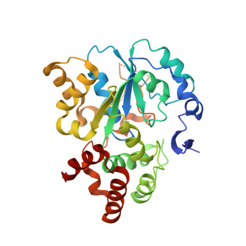 | |
UniProt | |||||
Find proteins for B7ZWN4 (Mus musculus) Explore B7ZWN4 Go to UniProtKB: B7ZWN4 | |||||
Entity Groups | |||||
| Sequence Clusters | 30% Identity50% Identity70% Identity90% Identity95% Identity100% Identity | ||||
| UniProt Group | B7ZWN4 | ||||
Sequence AnnotationsExpand | |||||
| |||||
| Ligands 3 Unique | |||||
|---|---|---|---|---|---|
| ID | Chains | Name / Formula / InChI Key | 2D Diagram | 3D Interactions | |
| A3P Query on A3P | BA [auth F] EA [auth G] HA [auth H] JA [auth I] LA [auth J] | ADENOSINE-3'-5'-DIPHOSPHATE C10 H15 N5 O10 P2 WHTCPDAXWFLDIH-KQYNXXCUSA-N |  | ||
| GOL Query on GOL | DA [auth F], OA [auth J] | GLYCEROL C3 H8 O3 PEDCQBHIVMGVHV-UHFFFAOYSA-N |  | ||
| CA Query on CA | AA [auth E] CA [auth F] FA [auth G] GA [auth G] IA [auth H] | CALCIUM ION Ca BHPQYMZQTOCNFJ-UHFFFAOYSA-N |  | ||
| Length ( Å ) | Angle ( ˚ ) |
|---|---|
| a = 166.174 | α = 90 |
| b = 81.127 | β = 119.95 |
| c = 166.18 | γ = 90 |
| Software Name | Purpose |
|---|---|
| REFMAC | refinement |
| HKL-2000 | data reduction |
| HKL-2000 | data scaling |
| REFMAC | phasing |
RCSB PDB (citation) is hosted by
RCSB PDB is a member of the

