5WUJ

Crystal structure of FliF-FliG complex from H. pylori


Experimental Data Snapshot

  • Method: X-RAY DIFFRACTION
  • Resolution: 2.30 Å
  • R-Value Free: 0.224 
  • R-Value Work: 0.188 
  • R-Value Observed: 0.190 

wwPDB Validation   3D Report Full Report


This is version 1.3 of the entry. See complete history


Literature

Crystal structure of the FliF-FliG complex from Helicobacter pylori yields insight into the assembly of the motor MS-C ring in the bacterial flagellum

Xue, C.Lam, K.H.Zhang, H.Sun, K.Lee, S.H.Chen, X.Au, S.W.N.

(2018) J Biol Chem 293: 2066-2078

  • DOI: https://doi.org/10.1074/jbc.M117.797936
  • Primary Citation of Related Structures:  
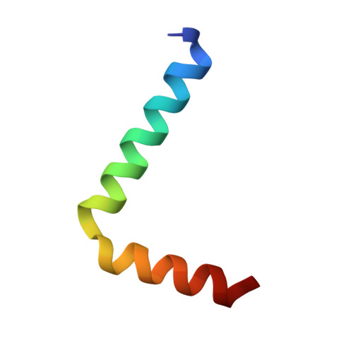
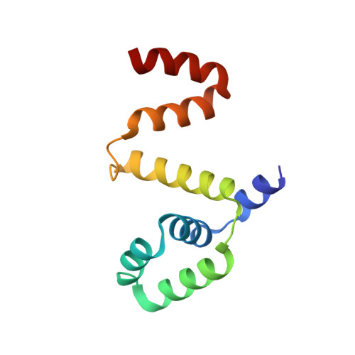
    5WUJ

  • PubMed Abstract: 

    The bacterial flagellar motor is a self-assembling supramolecular nanodevice. Its spontaneous biosynthesis is initiated by the insertion of the MS ring protein FliF into the inner membrane, followed by attachment of the switch protein FliG. Assembly of this multiprotein complex is tightly regulated to avoid nonspecific aggregation, but the molecular mechanisms governing flagellar assembly are unclear. Here, we present the crystal structure of the cytoplasmic domain of FliF complexed with the N-terminal domain of FliG (FliF C -FliG N ) from the bacterium Helicobacter pylori Within this complex, FliF C interacted with FliG N through extensive hydrophobic contacts similar to those observed in the FliF C -FliG N structure from the thermophile Thermotoga maritima , indicating conservation of the FliF C -FliG N interaction across bacterial species. Analysis of the crystal lattice revealed that the heterodimeric complex packs as a linear superhelix via stacking of the armadillo repeat-like motifs (ARM) of FliG N Notably, this linear helix was similar to that observed for the assembly of the FliG middle domain. We validated the in vivo relevance of the FliG N stacking by complementation studies in Escherichia coli Furthermore, structural comparison with apo FliG from the thermophile Aquifex aeolicus indicated that FliF regulates the conformational transition of FliG and exposes the complementary ARM-like motifs of FliG N , containing conserved hydrophobic residues. FliF apparently both provides a template for FliG polymerization and spatiotemporally controls subunit interactions within FliG. Our findings reveal that a small protein fold can serve as a versatile building block to assemble into a multiprotein machinery of distinct shapes for specific functions.


  • Organizational Affiliation

    From the Center for Protein Science and Crystallography, School of Life Sciences, The Chinese University of Hong Kong, Shatin, Hong Kong, China.


Macromolecules
Find similar proteins by:  (by identity cutoff)  |  3D Structure
Entity ID: 1
MoleculeChains Sequence LengthOrganismDetailsImage
Flagellar M-ring protein37Helicobacter pylori 26695Mutation(s): 0 
Gene Names: HP_0351
UniProt
Find proteins for O25118 (Helicobacter pylori (strain ATCC 700392 / 26695))
Explore O25118 
Go to UniProtKB:  O25118
Entity Groups  
Sequence Clusters30% Identity50% Identity70% Identity90% Identity95% Identity100% Identity
UniProt GroupO25118
Sequence Annotations
Expand
  • Reference Sequence
Find similar proteins by:  (by identity cutoff)  |  3D Structure
Entity ID: 2
MoleculeChains Sequence LengthOrganismDetailsImage
Flagellar motor switch protein FliG105Helicobacter pylori 26695Mutation(s): 0 
Gene Names: fliG
UniProt
Find proteins for O25119 (Helicobacter pylori (strain ATCC 700392 / 26695))
Explore O25119 
Go to UniProtKB:  O25119
Entity Groups  
Sequence Clusters30% Identity50% Identity70% Identity90% Identity95% Identity100% Identity
UniProt GroupO25119
Sequence Annotations
Expand
  • Reference Sequence
Small Molecules
Ligands 1 Unique
IDChains Name / Formula / InChI Key2D Diagram3D Interactions
GOL
Query on GOL

Download Ideal Coordinates CCD File 
C [auth B]GLYCEROL
C3 H8 O3
PEDCQBHIVMGVHV-UHFFFAOYSA-N
Experimental Data & Validation

Experimental Data

  • Method: X-RAY DIFFRACTION
  • Resolution: 2.30 Å
  • R-Value Free: 0.224 
  • R-Value Work: 0.188 
  • R-Value Observed: 0.190 
  • Space Group: P 32 2 1
Unit Cell:
Length ( Å )Angle ( ˚ )
a = 61.098α = 90
b = 61.098β = 90
c = 86.93γ = 120
Software Package:
Software NamePurpose
MOSFLMdata collection
SCALAdata scaling
PHENIXrefinement
PDB_EXTRACTdata extraction
MOSFLMdata reduction
AutoSolphasing

Structure Validation

View Full Validation Report


Entry History & Funding Information

Deposition Data


Funding OrganizationLocationGrant Number
GRFHong KongGRF 460112

Revision History  (Full details and data files)

  • Version 1.0: 2017-12-20
    Type: Initial release
  • Version 1.1: 2018-01-03
    Changes: Database references
  • Version 1.2: 2018-04-04
    Changes: Data collection, Database references
  • Version 1.3: 2024-03-20
    Changes: Data collection, Database references