5WTQ

Crystal structure of human proteasome-assembling chaperone PAC4


Experimental Data Snapshot

  • Method: X-RAY DIFFRACTION
  • Resolution: 1.90 Å
  • R-Value Free: 0.254 
  • R-Value Work: 0.204 
  • R-Value Observed: 0.207 

wwPDB Validation   3D Report Full Report


This is version 1.2 of the entry. See complete history


Literature

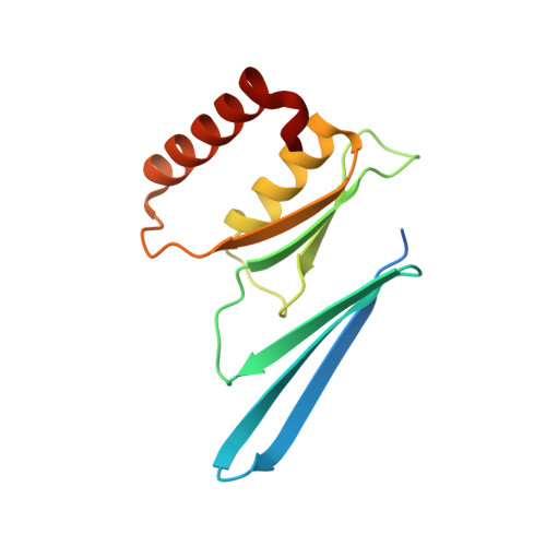
Crystal structure of human proteasome assembly chaperone PAC4 involved in proteasome formation

Kurimoto, E.Satoh, T.Ito, Y.Ishihara, E.Okamoto, K.Yagi-Utsumi, M.Tanaka, K.Kato, K.

(2017) Protein Sci 26: 1080-1085

  • DOI: https://doi.org/10.1002/pro.3153
  • Primary Citation of Related Structures:  
    5WTQ

  • PubMed Abstract: 

    The 26S proteasome is a large protein complex, responsible for degradation of ubiquinated proteins in eukaryotic cells. Eukaryotic proteasome formation is a highly ordered process that is assisted by several assembly chaperones. The assembly of its catalytic 20S core particle depends on at least five proteasome-specific chaperones, i.e., proteasome-assembling chaperons 1-4 (PAC1-4) and proteasome maturation protein (POMP). The orthologues of yeast assembly chaperones have been structurally characterized, whereas most mammalian assembly chaperones are not. In the present study, we determined a crystal structure of human PAC4 at 1.90-Å resolution. Our crystallographic data identify a hydrophobic surface that is surrounded by charged residues. The hydrophobic surface is complementary to that of its binding partner, PAC3. The surface also exhibits charge complementarity with the proteasomal α4-5 subunits. This will provide insights into human proteasome-assembling chaperones as potential anticancer drug targets.


  • Organizational Affiliation

    Faculty of Pharmacy, Meijo University, Tempaku-ku, Nagoya, 468-8503, Japan.


Macromolecules
Find similar proteins by:  (by identity cutoff)  |  3D Structure
Entity ID: 1
MoleculeChains Sequence LengthOrganismDetailsImage
Proteasome assembly chaperone 4
A, B, C, D
126Homo sapiensMutation(s): 1 
Gene Names: PSMG4C6orf86PAC4
UniProt & NIH Common Fund Data Resources
Find proteins for Q5JS54 (Homo sapiens)
Explore Q5JS54 
Go to UniProtKB:  Q5JS54
PHAROS:  Q5JS54
GTEx:  ENSG00000180822 
Entity Groups  
Sequence Clusters30% Identity50% Identity70% Identity90% Identity95% Identity100% Identity
UniProt GroupQ5JS54
Sequence Annotations
Expand
  • Reference Sequence
Experimental Data & Validation

Experimental Data

  • Method: X-RAY DIFFRACTION
  • Resolution: 1.90 Å
  • R-Value Free: 0.254 
  • R-Value Work: 0.204 
  • R-Value Observed: 0.207 
  • Space Group: F 2 2 2
Unit Cell:
Length ( Å )Angle ( ˚ )
a = 72.548α = 90
b = 152.779β = 90
c = 212.911γ = 90
Software Package:
Software NamePurpose
REFMACrefinement
HKL-2000data reduction
SCALEPACKdata scaling
PHENIXphasing

Structure Validation

View Full Validation Report


Entry History & Funding Information

Deposition Data


Funding OrganizationLocationGrant Number
JSPS KAKENHIJapan15H02491, 25102008

Revision History  (Full details and data files)

  • Version 1.0: 2017-03-22
    Type: Initial release
  • Version 1.1: 2017-05-24
    Changes: Database references
  • Version 1.2: 2024-03-20
    Changes: Data collection, Database references, Derived calculations, Refinement description