5WTL

Crystal structure of the periplasmic portion of outer membrane protein A (OmpA) from Capnocytophaga gingivalis


Experimental Data Snapshot

  • Method: X-RAY DIFFRACTION
  • Resolution: 2.30 Å
  • R-Value Free: 0.255 
  • R-Value Work: 0.203 
  • R-Value Observed: 0.206 

wwPDB Validation   3D Report Full Report


This is version 1.2 of the entry. See complete history


Literature

Structure of thrombospondin type 3 repeats in bacterial outer membrane protein A reveals its intra-repeat disulfide bond-dependent calcium-binding capability.

Dai, S.Sun, C.Tan, K.Ye, S.Zhang, R.

(2017) Cell Calcium 66: 78-89

  • DOI: https://doi.org/10.1016/j.ceca.2017.05.016
  • Primary Citation of Related Structures:  
    5WTL, 5WTP

  • PubMed Abstract: 

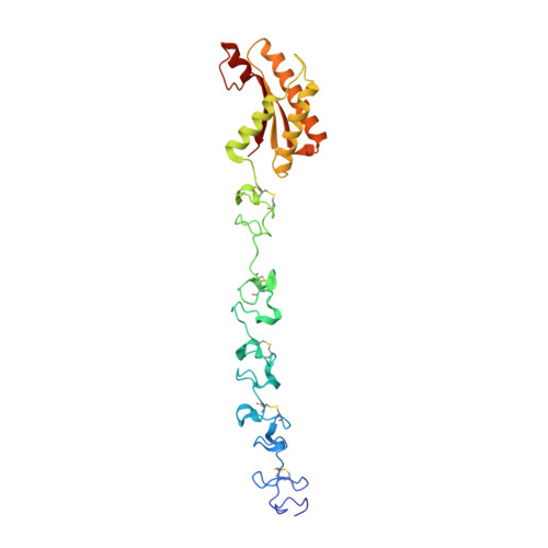
    Eukaryotic thrombospondin type 3 repeat (TT3R) is an efficient calcium ion (Ca 2+ ) binding motif only found in mammalian thrombospondin family. TT3R has also been found in prokaryotic cellulase Cel5G, which was thought to forfeit the Ca 2+ -binding capability due to the formation of intra-repeat disulfide bonds, instead of the inter-repeat ones possessed by eukaryotic TT3Rs. In this study, we have identified an enormous number of prokaryotic TT3R-containing proteins belonging to several different protein families, including outer membrane protein A (OmpA), an important structural protein connecting the outer membrane and the periplasmic peptidoglycan layer in gram-negative bacteria. Here, we report the crystal structure of the periplasmic region of OmpA from Capnocytophaga gingivalis, which contains a linker region comprising five consecutive TT3Rs. The structure of OmpA-TT3R exhibits a well-ordered architecture organized around two tightly-coordinated Ca 2+ and confirms the presence of abnormal intra-repeat disulfide bonds. Further mutagenesis studies showed that the Ca 2+ -binding capability of OmpA-TT3R is indeed dependent on the proper formation of intra-repeat disulfide bonds, which help to fix a conserved glycine residue at its proper position for Ca 2+ coordination. Additionally, despite lacking inter-repeat disulfide bonds, the interfaces between adjacent OmpA-TT3Rs are enhanced by both hydrophobic and conserved aromatic-proline interactions.


  • Organizational Affiliation

    National Laboratory of Biomacromolecules, Institute of Biophysics, Chinese Academy of Sciences, Beijing 100101, People's Republic of China.


Macromolecules
Find similar proteins by:  (by identity cutoff)  |  3D Structure
Entity ID: 1
MoleculeChains Sequence LengthOrganismDetailsImage
OmpA family protein
A, B, C, D
261Capnocytophaga gingivalis ATCC 33624Mutation(s): 0 
Gene Names: CAPGI0001_0903
UniProt
Find proteins for C2M2E7 (Capnocytophaga gingivalis)
Explore C2M2E7 
Go to UniProtKB:  C2M2E7
Entity Groups  
Sequence Clusters30% Identity50% Identity70% Identity90% Identity95% Identity100% Identity
UniProt GroupC2M2E7
Sequence Annotations
Expand
  • Reference Sequence
Small Molecules
Ligands 3 Unique
IDChains Name / Formula / InChI Key2D Diagram3D Interactions
MES
Query on MES

Download Ideal Coordinates CCD File 
KA [auth C]2-(N-MORPHOLINO)-ETHANESULFONIC ACID
C6 H13 N O4 S
SXGZJKUKBWWHRA-UHFFFAOYSA-N
SO4
Query on SO4

Download Ideal Coordinates CCD File 
LA [auth C],
Z [auth B]
SULFATE ION
O4 S
QAOWNCQODCNURD-UHFFFAOYSA-L
CA
Query on CA

Download Ideal Coordinates CCD File 
AA [auth C]
BA [auth C]
CA [auth C]
DA [auth C]
E [auth A]
AA [auth C],
BA [auth C],
CA [auth C],
DA [auth C],
E [auth A],
EA [auth C],
F [auth A],
FA [auth C],
G [auth A],
GA [auth C],
H [auth A],
HA [auth C],
I [auth A],
IA [auth C],
J [auth A],
JA [auth C],
K [auth A],
L [auth A],
M [auth A],
MA [auth C],
N [auth A],
NA [auth D],
O [auth A],
OA [auth D],
P [auth B],
PA [auth D],
Q [auth B],
QA [auth D],
R [auth B],
RA [auth D],
S [auth B],
SA [auth D],
T [auth B],
TA [auth D],
U [auth B],
UA [auth D],
V [auth B],
VA [auth D],
W [auth B],
X [auth B],
Y [auth B]
CALCIUM ION
Ca
BHPQYMZQTOCNFJ-UHFFFAOYSA-N
Experimental Data & Validation

Experimental Data

  • Method: X-RAY DIFFRACTION
  • Resolution: 2.30 Å
  • R-Value Free: 0.255 
  • R-Value Work: 0.203 
  • R-Value Observed: 0.206 
  • Space Group: P 21 21 21
Unit Cell:
Length ( Å )Angle ( ˚ )
a = 73.205α = 90
b = 87.343β = 90
c = 185.458γ = 90
Software Package:
Software NamePurpose
PHENIXrefinement
HKL-3000data reduction
HKL-3000data scaling
PHENIXphasing

Structure Validation

View Full Validation Report


Entry History 

Deposition Data

Revision History  (Full details and data files)

  • Version 1.0: 2017-12-13
    Type: Initial release
  • Version 1.1: 2018-09-12
    Changes: Data collection, Database references
  • Version 1.2: 2023-11-08
    Changes: Data collection, Database references, Derived calculations, Refinement description