5WRS

Crystal Structure of Fam20A in complex with ATP


Experimental Data Snapshot

  • Method: X-RAY DIFFRACTION
  • Resolution: 2.75 Å
  • R-Value Free: 0.238 
  • R-Value Work: 0.203 
  • R-Value Observed: 0.204 

wwPDB Validation   3D Report Full Report


Ligand Structure Quality Assessment 


This is version 2.1 of the entry. See complete history


Literature

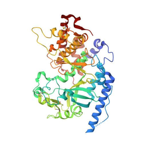
Structure of Fam20A reveals a pseudokinase featuring a unique disulfide pattern and inverted ATP-binding

Cui, J.Zhu, Q.Zhang, H.Cianfrocco, M.A.Leschziner, A.E.Dixon, J.E.Xiao, J.

(2017) Elife 6

  • DOI: https://doi.org/10.7554/eLife.23990
  • Primary Citation of Related Structures:  
    5WRR, 5WRS

  • PubMed Abstract: 

    Mutations in FAM20A cause tooth enamel defects known as Amelogenesis Imperfecta (AI) and renal calcification. We previously showed that Fam20A is a secretory pathway pseudokinase and allosterically activates the physiological casein kinase Fam20C to phosphorylate secreted proteins important for biomineralization (Cui et al., 2015). Here we report the nucleotide-free and ATP-bound structures of Fam20A. Fam20A exhibits a distinct disulfide bond pattern mediated by a unique insertion region. Loss of this insertion due to abnormal mRNA splicing interferes with the structure and function of Fam20A, resulting in AI. Fam20A binds ATP in the absence of divalent cations, and strikingly, ATP is bound in an inverted orientation compared to other kinases. Fam20A forms a dimer in the crystal, and residues in the dimer interface are critical for Fam20C activation. Together, these results provide structural insights into the function of Fam20A and shed light on the mechanism by which Fam20A mutations cause disease.


  • Organizational Affiliation

    Department of Pharmacology, University of California, San Diego, United States.


Macromolecules
Find similar proteins by:  (by identity cutoff)  |  3D Structure
Entity ID: 1
MoleculeChains Sequence LengthOrganismDetailsImage
Pseudokinase FAM20A
A, B
438Homo sapiensMutation(s): 1 
Gene Names: FAM20AUNQ9388/PRO34279
UniProt & NIH Common Fund Data Resources
Find proteins for Q96MK3 (Homo sapiens)
Explore Q96MK3 
Go to UniProtKB:  Q96MK3
PHAROS:  Q96MK3
GTEx:  ENSG00000108950 
Entity Groups  
Sequence Clusters30% Identity50% Identity70% Identity90% Identity95% Identity100% Identity
UniProt GroupQ96MK3
Sequence Annotations
Expand
  • Reference Sequence
Oligosaccharides

Help

Entity ID: 2
MoleculeChains Length2D Diagram Glycosylation3D Interactions
2-acetamido-2-deoxy-beta-D-glucopyranose-(1-4)-2-acetamido-2-deoxy-beta-D-glucopyranose
C, D
2N-Glycosylation
Glycosylation Resources
GlyTouCan:  G42666HT
GlyCosmos:  G42666HT
GlyGen:  G42666HT
Experimental Data & Validation

Experimental Data

  • Method: X-RAY DIFFRACTION
  • Resolution: 2.75 Å
  • R-Value Free: 0.238 
  • R-Value Work: 0.203 
  • R-Value Observed: 0.204 
  • Space Group: P 32 2 1
Unit Cell:
Length ( Å )Angle ( ˚ )
a = 157.202α = 90
b = 157.202β = 90
c = 144.802γ = 120
Software Package:
Software NamePurpose
PHENIXrefinement
HKL-2000data scaling
PDB_EXTRACTdata extraction
HKL-2000data reduction
PHASERphasing
SCALEPACKdata scaling

Structure Validation

View Full Validation Report


Ligand Structure Quality Assessment 


Entry History 

Deposition Data

  • Released Date: 2017-05-03 
  • Deposition Author(s): Zhu, Q.

Revision History  (Full details and data files)

  • Version 1.0: 2017-05-03
    Type: Initial release
  • Version 1.1: 2017-05-24
    Changes: Database references
  • Version 2.0: 2020-07-29
    Type: Remediation
    Reason: Carbohydrate remediation
    Changes: Advisory, Atomic model, Data collection, Derived calculations, Structure summary
  • Version 2.1: 2023-11-08
    Changes: Data collection, Database references, Refinement description, Structure summary