5WPN

Zn-bound Structure of Chaetopterus variopedatus Ferritin


Experimental Data Snapshot

  • Method: X-RAY DIFFRACTION
  • Resolution: 1.57 Å
  • R-Value Free: 0.163 
  • R-Value Work: 0.138 
  • R-Value Observed: 0.139 

wwPDB Validation   3D Report Full Report


This is version 1.3 of the entry. See complete history


Literature

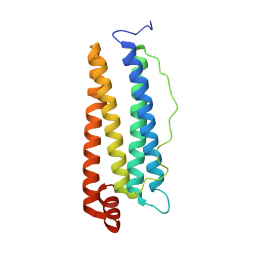
First biochemical and crystallographic characterization of a fast-performing ferritin from a marine invertebrate.

De Meulenaere, E.Bailey, J.B.Tezcan, F.A.Deheyn, D.D.

(2017) Biochem J 474: 4193-4206

  • DOI: https://doi.org/10.1042/BCJ20170681
  • Primary Citation of Related Structures:  
    5WPN

  • PubMed Abstract: 

    Ferritin, a multimeric cage-like enzyme, is integral to iron metabolism across all phyla through the sequestration and storage of iron through efficient ferroxidase activity. While ferritin sequences from ∼900 species have been identified, crystal structures from only 50 species have been reported, the majority from bacterial origin. We recently isolated a secreted ferritin from the marine invertebrate Chaetopterus sp. (parchment tube worm), which resides in muddy coastal seafloors. Here, we present the first ferritin from a marine invertebrate to be crystallized and its biochemical characterization. The initial ferroxidase reaction rate of recombinant Chaetopterus ferritin (ChF) is 8-fold faster than that of recombinant human heavy-chain ferritin (HuHF). To our knowledge, this protein exhibits the fastest catalytic performance ever described for a ferritin variant. In addition to the high-velocity ferroxidase activity, ChF is unique in that it is secreted by Chaetopterus in a bioluminescent mucus. Previous work has linked the availability of Fe 2+ to this long-lived bioluminescence, suggesting a potential function for the secreted ferritin. Comparative biochemical analyses indicated that both ChF and HuHF showed similar behavior toward changes in pH, temperature, and salt concentration. Comparison of their crystal structures shows no significant differences in the catalytic sites. Notable differences were found in the residues that line both 3-fold and 4-fold pores, potentially leading to increased flexibility, reduced steric hindrance, or a more efficient pathway for Fe 2+ transportation to the ferroxidase site. These suggested residues could contribute to the understanding of iron translocation through the ferritin shell to the ferroxidase site.


  • Organizational Affiliation

    Marine Biology Research Division, Scripps Institution of Oceanography, University of California, San Diego, La Jolla, CA 92037, U.S.A.


Macromolecules
Find similar proteins by:  (by identity cutoff)  |  3D Structure
Entity ID: 1
MoleculeChains Sequence LengthOrganismDetailsImage
Ferritin171Chaetopterus variopedatusMutation(s): 1 
EC: 1.16.3.1
UniProt
Find proteins for A0A075ML49 (Chaetopterus variopedatus)
Explore A0A075ML49 
Go to UniProtKB:  A0A075ML49
Entity Groups  
Sequence Clusters30% Identity50% Identity70% Identity90% Identity95% Identity100% Identity
UniProt GroupA0A075ML49
Sequence Annotations
Expand
  • Reference Sequence
Small Molecules
Ligands 5 Unique
IDChains Name / Formula / InChI Key2D Diagram3D Interactions
PEG
Query on PEG

Download Ideal Coordinates CCD File 
P [auth A],
Q [auth A]
DI(HYDROXYETHYL)ETHER
C4 H10 O3
MTHSVFCYNBDYFN-UHFFFAOYSA-N
ZN
Query on ZN

Download Ideal Coordinates CCD File 
B [auth A]
C [auth A]
D [auth A]
E [auth A]
F [auth A]
B [auth A],
C [auth A],
D [auth A],
E [auth A],
F [auth A],
G [auth A],
H [auth A],
I [auth A],
J [auth A],
K [auth A],
L [auth A],
M [auth A]
ZINC ION
Zn
PTFCDOFLOPIGGS-UHFFFAOYSA-N
EDO
Query on EDO

Download Ideal Coordinates CCD File 
R [auth A]1,2-ETHANEDIOL
C2 H6 O2
LYCAIKOWRPUZTN-UHFFFAOYSA-N
CA
Query on CA

Download Ideal Coordinates CCD File 
N [auth A]CALCIUM ION
Ca
BHPQYMZQTOCNFJ-UHFFFAOYSA-N
CL
Query on CL

Download Ideal Coordinates CCD File 
O [auth A]CHLORIDE ION
Cl
VEXZGXHMUGYJMC-UHFFFAOYSA-M
Experimental Data & Validation

Experimental Data

  • Method: X-RAY DIFFRACTION
  • Resolution: 1.57 Å
  • R-Value Free: 0.163 
  • R-Value Work: 0.138 
  • R-Value Observed: 0.139 
  • Space Group: F 4 3 2
Unit Cell:
Length ( Å )Angle ( ˚ )
a = 181.87α = 90
b = 181.87β = 90
c = 181.87γ = 90
Software Package:
Software NamePurpose
PHENIXrefinement
iMOSFLMdata reduction
Aimlessdata scaling
PHASERphasing

Structure Validation

View Full Validation Report


Entry History 

Deposition Data

Revision History  (Full details and data files)

  • Version 1.0: 2017-12-06
    Type: Initial release
  • Version 1.1: 2018-01-03
    Changes: Database references
  • Version 1.2: 2024-03-13
    Changes: Data collection, Database references, Derived calculations
  • Version 1.3: 2024-04-03
    Changes: Refinement description