5WMM

Crystal structure of an adenylation domain interrupted by a methylation domain (AMA4) from nonribosomal peptide synthetase TioS


Experimental Data Snapshot

  • Method: X-RAY DIFFRACTION
  • Resolution: 2.90 Å
  • R-Value Free: 0.253 
  • R-Value Work: 0.236 
  • R-Value Observed: 0.237 

wwPDB Validation   3D Report Full Report


Ligand Structure Quality Assessment 


This is version 1.3 of the entry. See complete history


Literature

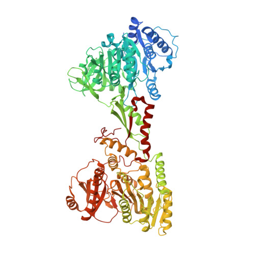
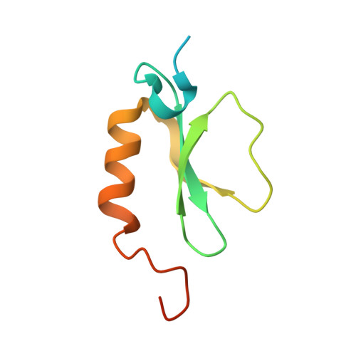
Structural basis for backbone N-methylation by an interrupted adenylation domain.

Mori, S.Pang, A.H.Lundy, T.A.Garzan, A.Tsodikov, O.V.Garneau-Tsodikova, S.

(2018) Nat Chem Biol 14: 428-430

  • DOI: https://doi.org/10.1038/s41589-018-0014-7
  • Primary Citation of Related Structures:  
    5WMM

  • PubMed Abstract: 

    Interrupted adenylation domains are enigmatic fusions, in which one enzyme is inserted into another to form a highly unusual bifunctional enzyme. We present the first crystal structure of an interrupted adenylation domain that reveals a unique embedded methyltransferase. The structure and functional data provide insight into how these enzymes N-methylate amino acid precursors en route to nonribosomal peptides.


  • Organizational Affiliation

    Department of Pharmaceutical Sciences, College of Pharmacy, University of Kentucky, Lexington, KY, USA.


Macromolecules
Find similar proteins by:  (by identity cutoff)  |  3D Structure
Entity ID: 1
MoleculeChains Sequence LengthOrganismDetailsImage
NRPS926Micromonospora sp. ML1Mutation(s): 0 
Gene Names: tioS
UniProt
Find proteins for Q333U7 (Micromonospora sp. ML1)
Explore Q333U7 
Go to UniProtKB:  Q333U7
Entity Groups  
Sequence Clusters30% Identity50% Identity70% Identity90% Identity95% Identity100% Identity
UniProt GroupQ333U7
Sequence Annotations
Expand
  • Reference Sequence
Find similar proteins by:  (by identity cutoff)  |  3D Structure
Entity ID: 2
MoleculeChains Sequence LengthOrganismDetailsImage
MbtH homologue87Micromonospora sp. ML1Mutation(s): 0 
Gene Names: tioT
UniProt
Find proteins for Q333U6 (Micromonospora sp. ML1)
Explore Q333U6 
Go to UniProtKB:  Q333U6
Entity Groups  
Sequence Clusters30% Identity50% Identity70% Identity90% Identity95% Identity100% Identity
UniProt GroupQ333U6
Sequence Annotations
Expand
  • Reference Sequence
Small Molecules
Ligands 4 Unique
IDChains Name / Formula / InChI Key2D Diagram3D Interactions
B6G
Query on B6G

Download Ideal Coordinates CCD File 
C [auth A](2S)-2-amino-3-methylbutanoyl (2S,3S,4R,5R)-5-(6-amino-9H-purin-9-yl)-3,4-dihydroxyoxolan-2-yl hydrogen (S)-phosphate
C14 H21 N6 O8 P
PJUFCHCQTYUTMG-JDUCOKCXSA-N
SAH
Query on SAH

Download Ideal Coordinates CCD File 
D [auth A]S-ADENOSYL-L-HOMOCYSTEINE
C14 H20 N6 O5 S
ZJUKTBDSGOFHSH-WFMPWKQPSA-N
CA
Query on CA

Download Ideal Coordinates CCD File 
H [auth A],
I [auth A],
J [auth A]
CALCIUM ION
Ca
BHPQYMZQTOCNFJ-UHFFFAOYSA-N
CL
Query on CL

Download Ideal Coordinates CCD File 
E [auth A],
F [auth A],
G [auth A]
CHLORIDE ION
Cl
VEXZGXHMUGYJMC-UHFFFAOYSA-M
Experimental Data & Validation

Experimental Data

  • Method: X-RAY DIFFRACTION
  • Resolution: 2.90 Å
  • R-Value Free: 0.253 
  • R-Value Work: 0.236 
  • R-Value Observed: 0.237 
  • Space Group: P 32 2 1
Unit Cell:
Length ( Å )Angle ( ˚ )
a = 136.712α = 90
b = 136.712β = 90
c = 228.234γ = 120
Software Package:
Software NamePurpose
REFMACrefinement
HKL-2000data reduction
HKL-2000data scaling
PHASERphasing

Structure Validation

View Full Validation Report


Ligand Structure Quality Assessment 


Entry History 

Deposition Data

Revision History  (Full details and data files)

  • Version 1.0: 2018-03-14
    Type: Initial release
  • Version 1.1: 2018-04-04
    Changes: Data collection, Database references
  • Version 1.2: 2018-04-25
    Changes: Data collection, Database references
  • Version 1.3: 2023-10-04
    Changes: Data collection, Database references, Refinement description