


5WKV
Structure of an acid sensing ion channel in a resting state with calcium
- PDB DOI: https://doi.org/10.2210/pdb5WKV/pdb
- Classification: TRANSPORT PROTEIN
- Organism(s): Gallus gallus
- Expression System: Homo sapiens
- Mutation(s): No
- Membrane Protein: Yes PDBTMMemProtMDmpstruc
- Deposited: 2017-07-25 Released: 2018-03-14
- Funding Organization(s): National Institutes of Health/National Institute of Diabetes and Digestive and Kidney Disease (NIH/NIDDK), National Institutes of Health/National Institute of Neurological Disorders and Stroke (NIH/NINDS)
Experimental Data Snapshot
- Method: X-RAY DIFFRACTION
- Resolution: 3.20 Å
- R-Value Free: 0.297
- R-Value Work: 0.287
- R-Value Observed: 0.287

This is version 2.1 of the entry. See complete history.

Macromolecules
Find similar proteins by:
(by identity cutoff) | 3D Structure
Entity ID: 1 | |||||
|---|---|---|---|---|---|
| Molecule | Chains | Sequence Length | Organism | Details | Image |
| Acid-sensing ion channel 1 | A, B [auth C], C [auth B] | 439 | Gallus gallus | Mutation(s): 0 Gene Names: ASIC1, ACCN2 Membrane Entity: Yes | 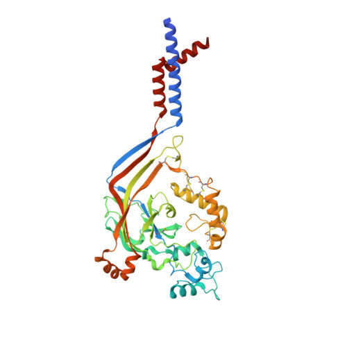 |
UniProt | |||||
Find proteins for Q1XA76 (Gallus gallus) Explore Q1XA76 Go to UniProtKB: Q1XA76 | |||||
Entity Groups | |||||
| Sequence Clusters | 30% Identity50% Identity70% Identity90% Identity95% Identity100% Identity | ||||
| UniProt Group | Q1XA76 | ||||
Sequence AnnotationsExpand | |||||
| |||||
Oligosaccharides

Small Molecules
| Ligands 3 Unique | |||||
|---|---|---|---|---|---|
| ID | Chains | Name / Formula / InChI Key | 2D Diagram | 3D Interactions | |
| NAG Query on NAG | F [auth A], G [auth A], K [auth C], N [auth B] | 2-acetamido-2-deoxy-beta-D-glucopyranose C8 H15 N O6 OVRNDRQMDRJTHS-FMDGEEDCSA-N |  | ||
| CA Query on CA | I [auth A] J [auth A] L [auth C] M [auth C] P [auth B] | CALCIUM ION Ca BHPQYMZQTOCNFJ-UHFFFAOYSA-N |  | ||
| CL Query on CL | H [auth A], O [auth B] | CHLORIDE ION Cl VEXZGXHMUGYJMC-UHFFFAOYSA-M |  | ||
Experimental Data & Validation
Experimental Data
- Method: X-RAY DIFFRACTION
- Resolution: 3.20 Å
- R-Value Free: 0.297
- R-Value Work: 0.287
- R-Value Observed: 0.287
- Space Group: P 21 21 21
Unit Cell:
| Length ( Å ) | Angle ( ˚ ) |
|---|---|
| a = 109.18 | α = 90 |
| b = 133.7 | β = 90 |
| c = 157.65 | γ = 90 |
| Software Name | Purpose |
|---|---|
| PHENIX | refinement |
| XDS | data reduction |
| XDS | data scaling |
| PHENIX | phasing |
Entry History & Funding Information
Deposition Data
- Released Date: 2018-03-14 Deposition Author(s): Yoder, N., Gouaux, E.
| Funding Organization | Location | Grant Number |
|---|---|---|
| National Institutes of Health/National Institute of Diabetes and Digestive and Kidney Disease (NIH/NIDDK) | United States | 5T32DK007680 |
| National Institutes of Health/National Institute of Neurological Disorders and Stroke (NIH/NINDS) | United States | 5F31NS096782 |
| National Institutes of Health/National Institute of Neurological Disorders and Stroke (NIH/NINDS) | United States | 5R01NS038631 |
Revision History (Full details and data files)
- Version 1.0: 2018-03-14
Type: Initial release - Version 1.1: 2018-03-21
Changes: Database references - Version 1.2: 2018-03-28
Changes: Data collection, Database references - Version 1.3: 2019-02-20
Changes: Author supporting evidence, Data collection - Version 1.4: 2019-12-18
Changes: Author supporting evidence - Version 2.0: 2020-07-29
Type: Remediation
Reason: Carbohydrate remediation
Changes: Advisory, Atomic model, Data collection, Derived calculations, Structure summary - Version 2.1: 2023-10-04
Changes: Data collection, Database references, Refinement description, Structure summary
