Structure of the Paramyxovirus Parainfluenza Virus 5 Nucleoprotein in Complex with an Amino-Terminal Peptide of the Phosphoprotein.
Aggarwal, M., Leser, G.P., Kors, C.A., Lamb, R.A.(2018) J Virol 92
- PubMed: 29237836
- DOI: https://doi.org/10.1128/JVI.01304-17
- Primary Citation of Related Structures:
5WKN - PubMed Abstract:
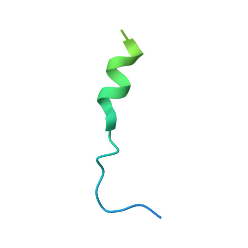
Parainfluenza virus 5 (PIV5) belongs to the family Paramyxoviridae , which consists of enveloped viruses with a nonsegmented negative-strand RNA genome encapsidated by the nucleoprotein (N). Paramyxovirus replication is regulated by the phosphoprotein (P) through protein-protein interactions with N and the RNA polymerase (L). The chaperone activity of P is essential to maintain the unassembled RNA-free form of N in order to prevent nonspecific RNA binding and premature N oligomerization. Here, we determined the crystal structure of unassembled PIV5 N in complex with a P peptide (N 0 P) derived from the N terminus of P (P50) at 2.65 Å. The PIV5 N 0 P consists of two domains: an N-terminal domain (NTD) and a C-terminal domain (CTD) separated by a hinge region. The cleft at the hinge region of RNA-bound PIV5 N was previously shown to be an RNA binding site. The N 0 P structure shows that the P peptide binds to the CTD of N and extends toward the RNA binding site to inhibit N oligomerization and, hence, RNA binding. Binding of P peptide also keeps the PIV5 N in the open form. A molecular dynamics (MD) analysis of both the open and closed forms of N shows the flexibility of the CTD and the preference of the N protein to be in an open conformation. The gradual opening of the hinge region, to release the RNA, was also observed. Together, these results advance our knowledge of the conformational swapping of N required for the highly regulated paramyxovirus replication. IMPORTANCE Paramyxovirus replication is regulated by the interaction of P with N and L proteins. Here, we report the crystal structure of unassembled parainfluenza virus 5 (PIV5) N chaperoned with P peptide. Our results provide a detailed understanding of the binding of P to N. The conformational switching of N between closed and open forms during its initial interaction with P, as well as during RNA release, was analyzed. Our data also show the plasticity of the CTD and the importance of domain movement for conformational switching. The results improve our understanding of the mechanism of interchanging N conformations for RNA replication and release.
Organizational Affiliation:
Department of Molecular Biosciences, Northwestern University, Evanston, Illinois, USA.