


Crystal structure of Pol III beta from Caulobacter crescentus
Dawes, F., Oakley, A.J.To be published.
Experimental Data Snapshot
wwPDB Validation 3D Report Full Report

Entity ID: 1 | |||||
|---|---|---|---|---|---|
| Molecule | Chains | Sequence Length | Organism | Details | Image |
| DNA polymerase III subunit beta | 379 | Caulobacter vibrioides CB15 | Mutation(s): 0 Gene Names: dnaN, CC_0156 EC: 2.7.7.7 | 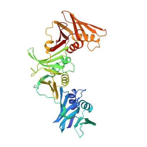 | |
UniProt | |||||
Find proteins for P0CAU5 (Caulobacter vibrioides (strain ATCC 19089 / CB15)) Explore P0CAU5 Go to UniProtKB: P0CAU5 | |||||
Entity Groups | |||||
| Sequence Clusters | 30% Identity50% Identity70% Identity90% Identity95% Identity100% Identity | ||||
| UniProt Group | P0CAU5 | ||||
Sequence AnnotationsExpand | |||||
| |||||
| Length ( Å ) | Angle ( ˚ ) |
|---|---|
| a = 83.855 | α = 90 |
| b = 59.572 | β = 92.89 |
| c = 86.49 | γ = 90 |
| Software Name | Purpose |
|---|---|
| REFMAC | refinement |
| DENZO | data reduction |
| SCALEPACK | data scaling |
| PHENIX | phasing |
RCSB PDB (citation) is hosted by
RCSB PDB is a member of the

