5W8S

Lipid A Disaccharide Synthase (LpxB)-7 solubilizing mutations


Experimental Data Snapshot

  • Method: X-RAY DIFFRACTION
  • Resolution: 2.10 Å
  • R-Value Free: 0.223 
  • R-Value Work: 0.195 
  • R-Value Observed: 0.197 

wwPDB Validation   3D Report Full Report


This is version 1.3 of the entry. See complete history


Literature

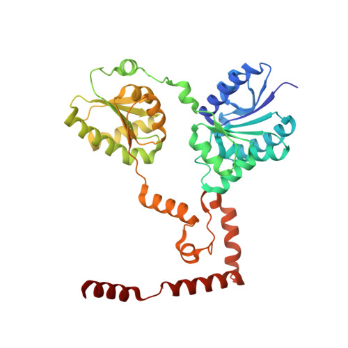
Crystal structure of lipid A disaccharide synthase LpxB from Escherichia coli.

Bohl, T.E.Shi, K.Lee, J.K.Aihara, H.

(2018) Nat Commun 9: 377-377

  • DOI: https://doi.org/10.1038/s41467-017-02712-9
  • Primary Citation of Related Structures:  
    5W8N, 5W8S, 5W8X

  • PubMed Abstract: 

    Most Gram-negative bacteria are surrounded by a glycolipid called lipopolysaccharide (LPS), which forms a barrier to hydrophobic toxins and, in pathogenic bacteria, is a virulence factor. During LPS biosynthesis, a membrane-associated glycosyltransferase (LpxB) forms a tetra-acylated disaccharide that is further acylated to form the membrane anchor moiety of LPS. Here we solve the structure of a soluble and catalytically competent LpxB by X-ray crystallography. The structure reveals that LpxB has a glycosyltransferase-B family fold but with a highly intertwined, C-terminally swapped dimer comprising four domains. We identify key catalytic residues with a product, UDP, bound in the active site, as well as clusters of hydrophobic residues that likely mediate productive membrane association or capture of lipidic substrates. These studies provide the basis for rational design of antibiotics targeting a crucial step in LPS biosynthesis.


  • Organizational Affiliation

    Department of Biochemistry, Molecular Biology, and Biophysics, University of Minnesota Twin Cities, Minneapolis, MN, 55455, USA.


Macromolecules
Find similar proteins by:  (by identity cutoff)  |  3D Structure
Entity ID: 1
MoleculeChains Sequence LengthOrganismDetailsImage
Lipid-A-disaccharide synthase382Escherichia coli BL21(DE3)Mutation(s): 7 
Gene Names: lpxBECBD_3437
EC: 2.4.1.182
UniProt
Find proteins for P10441 (Escherichia coli (strain K12))
Explore P10441 
Go to UniProtKB:  P10441
Entity Groups  
Sequence Clusters30% Identity50% Identity70% Identity90% Identity95% Identity100% Identity
UniProt GroupP10441
Sequence Annotations
Expand
  • Reference Sequence
Experimental Data & Validation

Experimental Data

  • Method: X-RAY DIFFRACTION
  • Resolution: 2.10 Å
  • R-Value Free: 0.223 
  • R-Value Work: 0.195 
  • R-Value Observed: 0.197 
  • Space Group: P 32 2 1
Unit Cell:
Length ( Å )Angle ( ˚ )
a = 68.016α = 90
b = 68.016β = 90
c = 155.139γ = 120
Software Package:
Software NamePurpose
PHENIXrefinement
XDSdata reduction
Cootmodel building
PHENIXphasing
Aimlessdata scaling

Structure Validation

View Full Validation Report


Entry History & Funding Information

Deposition Data


Funding OrganizationLocationGrant Number
National Institutes of Health/National Institute of General Medical Sciences (NIH/NIGMS)United StatesGM118047
National Institutes of Health/National Institute of General Medical Sciences (NIH/NIGMS)United StatesP41-GM103403
Department of Energy (DOE, United States)United StatesDE-AC02-06CH11357

Revision History  (Full details and data files)

  • Version 1.0: 2018-01-31
    Type: Initial release
  • Version 1.1: 2018-02-07
    Changes: Database references
  • Version 1.2: 2020-01-01
    Changes: Author supporting evidence
  • Version 1.3: 2024-03-13
    Changes: Data collection, Database references