5W6H

Crystal structure of Bacteriophage CBA120 tailspike protein 4 enzymatically active domain (TSP4dN, orf213)


Experimental Data Snapshot

  • Method: X-RAY DIFFRACTION
  • Resolution: 2.29 Å
  • R-Value Free: 0.170 
  • R-Value Work: 0.140 
  • R-Value Observed: 0.141 

wwPDB Validation   3D Report Full Report


This is version 1.2 of the entry. See complete history


Literature

Structure and Function of the Branched Receptor-Binding Complex of Bacteriophage CBA120.

Plattner, M.Shneider, M.M.Arbatsky, N.P.Shashkov, A.S.Chizhov, A.O.Nazarov, S.Prokhorov, N.S.Taylor, N.M.I.Buth, S.A.Gambino, M.Gencay, Y.E.Brondsted, L.Kutter, E.M.Knirel, Y.A.Leiman, P.G.

(2019) J Mol Biol 431: 3718-3739

  • DOI: https://doi.org/10.1016/j.jmb.2019.07.022
  • Primary Citation of Related Structures:  
    5W6F, 5W6H, 5W6P

  • PubMed Abstract: 

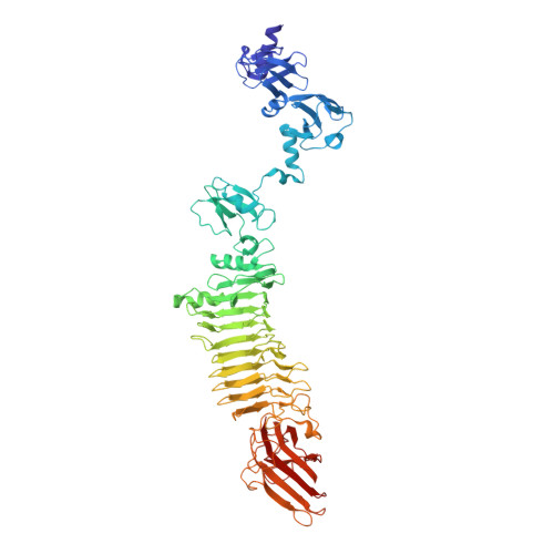
    Bacteriophages recognize their host cells with the help of tail fiber and tailspike proteins that bind, cleave, or modify certain structures on the cell surface. The spectrum of ligands to which the tail fibers and tailspikes can bind is the primary determinant of the host range. Bacteriophages with multiple tailspike/tail fibers are thought to have a wider host range than their less endowed relatives but the function of these proteins remains poorly understood. Here, we describe the structure, function, and substrate specificity of three tailspike proteins of bacteriophage CBA120-TSP2, TSP3 and TSP4 (orf211 through orf213, respectively). We show that tailspikes TSP2, TSP3 and TSP4 are hydrolases that digest the O157, O77, and O78 Escherichia coli O-antigens, respectively. We demonstrate that recognition of the E. coli O157:H7 host by CBA120 involves binding to and digesting the O157 O-antigen by TSP2. We report the crystal structure of TSP2 in complex with a repeating unit of the O157 O-antigen. We demonstrate that according to the specificity of its tailspikes TSP2, TSP3, and TSP4, CBA120 can infect E. coli O157, O77, and O78, respectively. We also show that CBA120 infects Salmonella enterica serovar Minnesota, and this host range expansion is likely due to the function of TSP1. Finally, we describe the assembly pathway and the architecture of the TSP1-TSP2-TSP3-TSP4 branched complex in CBA120 and its related ViI-like phages.


  • Organizational Affiliation

    Department of Biochemistry and Molecular Biology, Sealy Center for Structural Biology and Molecular Biophysics, University of Texas Medical Branch, 301 University Blvd, Galveston, TX 77555-0647, USA; École Polytechnique Fédérale de Lausanne, Lausanne CH-1015, Switzerland.


Macromolecules
Find similar proteins by:  (by identity cutoff)  |  3D Structure
Entity ID: 1
MoleculeChains Sequence LengthOrganismDetailsImage
tailspike protein 4
A, B, C
697Kuttervirus CBA120Mutation(s): 1 
Gene Names: orf213
UniProt
Find proteins for G3M192 (Escherichia phage Cba120)
Explore G3M192 
Go to UniProtKB:  G3M192
Entity Groups  
Sequence Clusters30% Identity50% Identity70% Identity90% Identity95% Identity100% Identity
UniProt GroupG3M192
Sequence Annotations
Expand
  • Reference Sequence
Small Molecules
Ligands 6 Unique
IDChains Name / Formula / InChI Key2D Diagram3D Interactions
SO4
Query on SO4

Download Ideal Coordinates CCD File 
GA [auth C]SULFATE ION
O4 S
QAOWNCQODCNURD-UHFFFAOYSA-L
ZN
Query on ZN

Download Ideal Coordinates CCD File 
P [auth A]ZINC ION
Zn
PTFCDOFLOPIGGS-UHFFFAOYSA-N
ACT
Query on ACT

Download Ideal Coordinates CCD File 
AA [auth C]
BA [auth C]
CA [auth C]
D [auth A]
DA [auth C]
AA [auth C],
BA [auth C],
CA [auth C],
D [auth A],
DA [auth C],
E [auth A],
EA [auth C],
F [auth A],
FA [auth C],
G [auth A],
H [auth A],
I [auth A],
J [auth A],
K [auth A],
L [auth A],
M [auth A],
Q [auth B],
R [auth B],
S [auth B],
T [auth B],
U [auth B],
V [auth B],
W [auth B],
X [auth B],
Y [auth C],
Z [auth C]
ACETATE ION
C2 H3 O2
QTBSBXVTEAMEQO-UHFFFAOYSA-M
K
Query on K

Download Ideal Coordinates CCD File 
N [auth A]POTASSIUM ION
K
NPYPAHLBTDXSSS-UHFFFAOYSA-N
CL
Query on CL

Download Ideal Coordinates CCD File 
HA [auth C]CHLORIDE ION
Cl
VEXZGXHMUGYJMC-UHFFFAOYSA-M
NA
Query on NA

Download Ideal Coordinates CCD File 
O [auth A]SODIUM ION
Na
FKNQFGJONOIPTF-UHFFFAOYSA-N
Experimental Data & Validation

Experimental Data

  • Method: X-RAY DIFFRACTION
  • Resolution: 2.29 Å
  • R-Value Free: 0.170 
  • R-Value Work: 0.140 
  • R-Value Observed: 0.141 
  • Space Group: P 41 2 2
Unit Cell:
Length ( Å )Angle ( ˚ )
a = 170.079α = 90
b = 170.079β = 90
c = 246.384γ = 90
Software Package:
Software NamePurpose
PHENIXrefinement
XDSdata reduction
XSCALEdata scaling
PHASERphasing

Structure Validation

View Full Validation Report


Entry History & Funding Information

Deposition Data


Funding OrganizationLocationGrant Number
EPFLSwitzerland0577-1

Revision History  (Full details and data files)

  • Version 1.0: 2018-10-24
    Type: Initial release
  • Version 1.1: 2019-11-06
    Changes: Data collection, Database references
  • Version 1.2: 2024-03-13
    Changes: Data collection, Database references, Derived calculations