5W4S

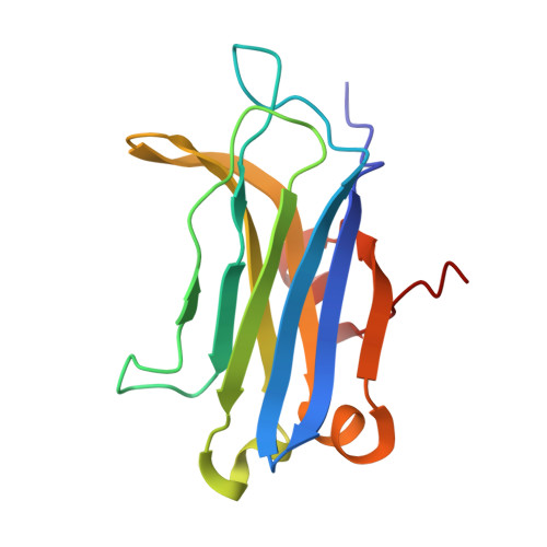
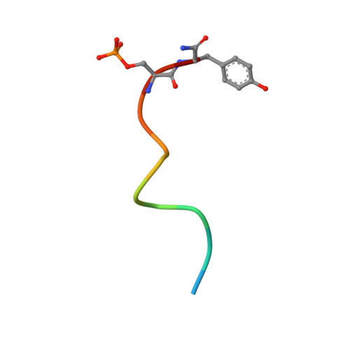
Solution structure of C2 domain from protein kinase C alpha in ternary complex with calcium and V5-pHM peptide

  • Classification: TRANSFERASE
  • Organism(s): Rattus norvegicus
  • Expression System: Escherichia coli
  • Mutation(s): No 

  • Deposited: 2017-06-12 Released: 2018-04-25 
  • Deposition Author(s): Yang, Y., Igumenova, T.I.
  • Funding Organization(s): Welch Foundation, National Institutes of Health/National Institute of General Medical Sciences (NIH/NIGMS), National Science Foundation (NSF, United States)

Experimental Data Snapshot

  • Method: SOLUTION NMR
  • Conformers Calculated: 400 
  • Conformers Submitted: 20 
  • Selection Criteria: structures with the lowest energy 

wwPDB Validation   3D Report Full Report


This is version 1.2 of the entry. See complete history


Literature

Structural Basis of Protein Kinase C alpha Regulation by the C-Terminal Tail.

Yang, Y.Shu, C.Li, P.Igumenova, T.I.

(2018) Biophys J 114: 1590-1603

  • DOI: https://doi.org/10.1016/j.bpj.2017.12.030
  • Primary Citation of Related Structures:  
    5W4S

  • PubMed Abstract: 

    Protein kinase C (PKC) isoenzymes are multi-modular proteins activated at the membrane surface to regulate signal transduction processes. When activated by second messengers, PKC undergoes a drastic conformational and spatial transition from the inactive cytosolic state to the activated membrane-bound state. The complete structure of either state of PKC remains elusive. We demonstrate, using NMR spectroscopy, that the isolated Ca 2+ -sensing membrane-binding C2 domain of the conventional PKCα interacts with a conserved hydrophobic motif of the kinase C-terminal region, and we report a structural model of the complex. Our data suggest that the C-terminal region plays a dual role in regulating the PKC activity: activating, through sensitization of PKC to intracellular Ca 2+ oscillations; and auto-inhibitory, through its interaction with a conserved positively charged region of the C2 domain.


  • Organizational Affiliation

    Department of Biochemistry and Biophysics, Texas A&M University, College Station, Texas.


Macromolecules
Find similar proteins by:  (by identity cutoff)  |  3D Structure
Entity ID: 1
MoleculeChains Sequence LengthOrganismDetailsImage
Protein kinase C alpha type139Rattus norvegicusMutation(s): 0 
Gene Names: PrkcaPkca
EC: 2.7.11.13
UniProt
Find proteins for P05696 (Rattus norvegicus)
Explore P05696 
Go to UniProtKB:  P05696
Entity Groups  
Sequence Clusters30% Identity50% Identity70% Identity90% Identity95% Identity100% Identity
UniProt GroupP05696
Sequence Annotations
Expand
  • Reference Sequence

Find similar proteins by:  Sequence   |   3D Structure  

Entity ID: 2
MoleculeChains Sequence LengthOrganismDetailsImage
V5-pHM peptide12Rattus norvegicusMutation(s): 0 
UniProt
Find proteins for P05696 (Rattus norvegicus)
Explore P05696 
Go to UniProtKB:  P05696
Entity Groups  
Sequence Clusters30% Identity50% Identity70% Identity90% Identity95% Identity100% Identity
UniProt GroupP05696
Sequence Annotations
Expand
  • Reference Sequence
Small Molecules
Ligands 1 Unique
IDChains Name / Formula / InChI Key2D Diagram3D Interactions
CA
Query on CA

Download Ideal Coordinates CCD File 
C [auth A],
D [auth A]
CALCIUM ION
Ca
BHPQYMZQTOCNFJ-UHFFFAOYSA-N
Modified Residues  1 Unique
IDChains TypeFormula2D DiagramParent
SEP
Query on SEP
B
L-PEPTIDE LINKINGC3 H8 N O6 PSER
Experimental Data & Validation

Experimental Data

  • Method: SOLUTION NMR
  • Conformers Calculated: 400 
  • Conformers Submitted: 20 
  • Selection Criteria: structures with the lowest energy 

Structure Validation

View Full Validation Report


Entry History & Funding Information

Deposition Data


Funding OrganizationLocationGrant Number
Welch FoundationUnited StatesA-1784
National Institutes of Health/National Institute of General Medical Sciences (NIH/NIGMS)United StatesGM108998
National Science Foundation (NSF, United States)United StatesCHE-1151435

Revision History  (Full details and data files)

  • Version 1.0: 2018-04-25
    Type: Initial release
  • Version 1.1: 2019-11-27
    Changes: Author supporting evidence, Data collection
  • Version 1.2: 2023-06-14
    Changes: Database references, Derived calculations, Other