5W3S

Cryo-electron microscopy structure of a TRPML3 ion channel


Experimental Data Snapshot

  • Method: ELECTRON MICROSCOPY
  • Resolution: 2.94 Å
  • Aggregation State: PARTICLE 
  • Reconstruction Method: SINGLE PARTICLE 

wwPDB Validation   3D Report Full Report


This is version 1.4 of the entry. See complete history


Literature

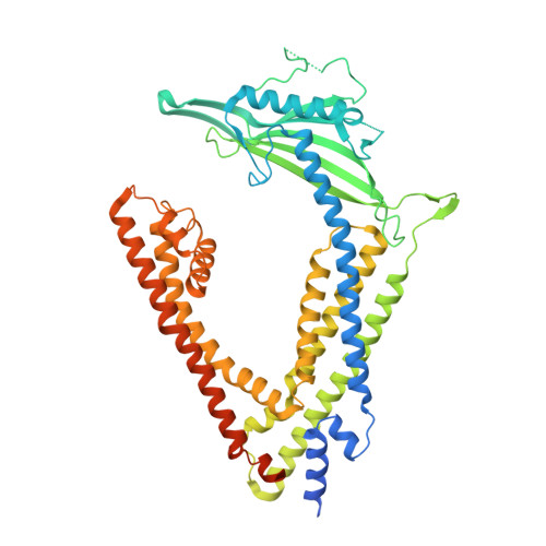
Cryo-electron microscopy structure of the lysosomal calcium-permeable channel TRPML3.

Hirschi, M.Herzik, M.A.Wie, J.Suo, Y.Borschel, W.F.Ren, D.Lander, G.C.Lee, S.Y.

(2017) Nature 550: 411-414

  • DOI: https://doi.org/10.1038/nature24055
  • Primary Citation of Related Structures:  
    5W3S

  • PubMed Abstract: 

    The modulation of ion channel activity by lipids is increasingly recognized as a fundamental component of cellular signalling. The transient receptor potential mucolipin (TRPML) channel family belongs to the TRP superfamily and is composed of three members: TRPML1-TRPML3. TRPMLs are the major Ca 2+ -permeable channels on late endosomes and lysosomes (LEL). They regulate the release of Ca 2+ from organelles, which is important for various physiological processes, including organelle trafficking and fusion. Loss-of-function mutations in the MCOLN1 gene, which encodes TRPML1, cause the neurodegenerative lysosomal storage disorder mucolipidosis type IV, and a gain-of-function mutation (Ala419Pro) in TRPML3 gives rise to the varitint-waddler (Va) mouse phenotype. Notably, TRPML channels are activated by the low-abundance and LEL-enriched signalling lipid phosphatidylinositol-3,5-bisphosphate (PtdIns(3,5)P 2 ), whereas other phosphoinositides such as PtdIns(4,5)P 2 , which is enriched in plasma membranes, inhibit TRPMLs. Conserved basic residues at the N terminus of the channel are important for activation by PtdIns(3,5)P 2 and inhibition by PtdIns(4,5)P 2 . However, owing to a lack of structural information, the mechanism by which TRPML channels recognize PtdIns(3,5)P 2 and increase their Ca 2+ conductance remains unclear. Here we present the cryo-electron microscopy (cryo-EM) structure of a full-length TRPML3 channel from the common marmoset (Callithrix jacchus) at an overall resolution of 2.9 Å. Our structure reveals not only the molecular basis of ion conduction but also the unique architecture of TRPMLs, wherein the voltage sensor-like domain is linked to the pore via a cytosolic domain that we term the mucolipin domain. Combined with functional studies, these data suggest that the mucolipin domain is responsible for PtdIns(3,5)P 2 binding and subsequent channel activation, and that it acts as a 'gating pulley' for lipid-dependent TRPML gating.


  • Organizational Affiliation

    Department of Biochemistry, Duke University School of Medicine, Durham, North Carolina 27710, USA.


Macromolecules
Find similar proteins by:  (by identity cutoff)  |  3D Structure
Entity ID: 1
MoleculeChains Sequence LengthOrganismDetailsImage
Mucolipin-3 isoform 1
A, B, C, D
561Callithrix jacchusMutation(s): 1 
Gene Names: MCOLN3
Membrane Entity: Yes 
UniProt
Find proteins for F6RG56 (Callithrix jacchus)
Explore F6RG56 
Go to UniProtKB:  F6RG56
Entity Groups  
Sequence Clusters30% Identity50% Identity70% Identity90% Identity95% Identity100% Identity
UniProt GroupF6RG56
Sequence Annotations
Expand
  • Reference Sequence
Small Molecules
Ligands 3 Unique
IDChains Name / Formula / InChI Key2D Diagram3D Interactions
3PE
Query on 3PE

Download Ideal Coordinates CCD File 
H [auth A],
M [auth B],
O [auth C],
X [auth D]
1,2-Distearoyl-sn-glycerophosphoethanolamine
C41 H82 N O8 P
LVNGJLRDBYCPGB-LDLOPFEMSA-N
Y01
Query on Y01

Download Ideal Coordinates CCD File 
E [auth A]
I [auth A]
J [auth A]
K [auth B]
L [auth B]
E [auth A],
I [auth A],
J [auth A],
K [auth B],
L [auth B],
N [auth B],
P [auth C],
T [auth C],
U [auth C],
W [auth D],
Y [auth D],
Z [auth D]
CHOLESTEROL HEMISUCCINATE
C31 H50 O4
WLNARFZDISHUGS-MIXBDBMTSA-N
NA
Query on NA

Download Ideal Coordinates CCD File 
F [auth A]
G [auth A]
Q [auth C]
R [auth C]
S [auth C]
F [auth A],
G [auth A],
Q [auth C],
R [auth C],
S [auth C],
V [auth D]
SODIUM ION
Na
FKNQFGJONOIPTF-UHFFFAOYSA-N
Experimental Data & Validation

Experimental Data

  • Method: ELECTRON MICROSCOPY
  • Resolution: 2.94 Å
  • Aggregation State: PARTICLE 
  • Reconstruction Method: SINGLE PARTICLE 

Structure Validation

View Full Validation Report


Entry History & Funding Information

Deposition Data


Funding OrganizationLocationGrant Number
National Institutes of Health/National Institute of Neurological Disorders and Stroke (NIH/NINDS)United StatesR35NS097241
National Institutes of Health/National Institute of Biomedical Imaging and Bioengineering (NIH/NIBIB)United StatesDP2EB020402

Revision History  (Full details and data files)

  • Version 1.0: 2017-10-11
    Type: Initial release
  • Version 1.1: 2017-10-25
    Changes: Database references
  • Version 1.2: 2017-11-01
    Changes: Database references
  • Version 1.3: 2019-12-11
    Changes: Author supporting evidence
  • Version 1.4: 2024-03-13
    Changes: Data collection, Database references, Derived calculations, Structure summary