5W3D

The structure of kinesin-14 wild-type Ncd-ADP dimer


Experimental Data Snapshot

  • Method: X-RAY DIFFRACTION
  • Resolution: 2.79 Å
  • R-Value Free: 0.280 
  • R-Value Work: 0.235 
  • R-Value Observed: 0.238 

wwPDB Validation   3D Report Full Report


Ligand Structure Quality Assessment 


This is version 1.1 of the entry. See complete history


Literature

Structural basis of small molecule ATPase inhibition of a human mitotic kinesin motor protein.

Park, H.W.Ma, Z.Zhu, H.Jiang, S.Robinson, R.C.Endow, S.A.

(2017) Sci Rep 7: 15121-15121

  • DOI: https://doi.org/10.1038/s41598-017-14754-6
  • Primary Citation of Related Structures:  
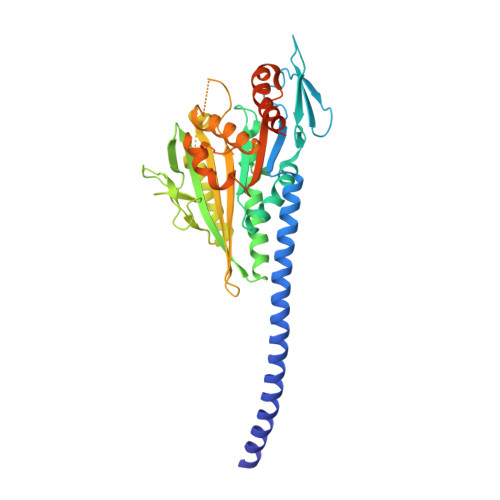
    5W3D, 5WDE, 5WDH

  • PubMed Abstract: 

    Kinesin microtubule motor proteins play essential roles in division, including attaching chromosomes to spindles and crosslinking microtubules for spindle assembly. Human kinesin-14 KIFC1 is unique in that cancer cells with amplified centrosomes are dependent on the motor for viable division because of its ability to cluster centrosomes and form bipolar spindles, but it is not required for division in almost all normal cells. Screens for small molecule inhibitors of KIFC1 have yielded several candidates for further development, but obtaining structural data to determine their sites of binding has been difficult. Here we compare a previously unreported KIFC1 crystal structure with new structures of two closely related kinesin-14 proteins, Ncd and KIFC3, to determine the potential binding site of a known KIFC1 ATPase inhibitor, AZ82. We analyze the previously identified kinesin inhibitor binding sites and identify features of AZ82 that favor binding to one of the sites, the α4/α6 site. This selectivity can be explained by unique structural features of the KIFC1 α4/α6 binding site. These features may help improve the drug-like properties of AZ82 and other specific KIFC1 inhibitors.


  • Organizational Affiliation

    Structural Genomics Consortium, University of Toronto, Toronto, ON M5G 1L7, Canada.


Macromolecules
Find similar proteins by:  (by identity cutoff)  |  3D Structure
Entity ID: 1
MoleculeChains Sequence LengthOrganismDetailsImage
Protein claret segregational
A, B
412Drosophila melanogasterMutation(s): 0 
Gene Names: ncdCA(ND)CG7831
UniProt
Find proteins for P20480 (Drosophila melanogaster)
Explore P20480 
Go to UniProtKB:  P20480
Entity Groups  
Sequence Clusters30% Identity50% Identity70% Identity90% Identity95% Identity100% Identity
UniProt GroupP20480
Sequence Annotations
Expand
  • Reference Sequence
Experimental Data & Validation

Experimental Data

  • Method: X-RAY DIFFRACTION
  • Resolution: 2.79 Å
  • R-Value Free: 0.280 
  • R-Value Work: 0.235 
  • R-Value Observed: 0.238 
  • Space Group: C 1 2 1
Unit Cell:
Length ( Å )Angle ( ˚ )
a = 163.271α = 90
b = 67.249β = 97.92
c = 94.421γ = 90
Software Package:
Software NamePurpose
REFMACrefinement
SCALAdata scaling
Cootmodel building
PDB_EXTRACTdata extraction
XDSdata reduction
MOLREPphasing

Structure Validation

View Full Validation Report


Ligand Structure Quality Assessment 


Entry History 

Deposition Data

Revision History  (Full details and data files)

  • Version 1.0: 2017-12-20
    Type: Initial release
  • Version 1.1: 2023-10-04
    Changes: Data collection, Database references, Derived calculations, Refinement description