5W1A

The first X-ray crystal structure of an insect muscle myosin. Drosophila melanogaster, skeletal muscle myosin II, an embryonic isoform, subfragment-1


Experimental Data Snapshot

  • Method: X-RAY DIFFRACTION
  • Resolution: 2.23 Å
  • R-Value Free: 0.220 
  • R-Value Work: 0.178 
  • R-Value Observed: 0.180 

wwPDB Validation   3D Report Full Report


This is version 1.3 of the entry. See complete history


Literature

X-ray crystallographic and molecular dynamic analyses of Drosophila melanogaster embryonic muscle myosin define domains responsible for isoform-specific properties.

Caldwell, J.T.Mermelstein, D.J.Walker, R.C.Bernstein, S.I.Huxford, T.

(2019) J Mol Biol 

  • DOI: https://doi.org/10.1016/j.jmb.2019.11.013
  • Primary Citation of Related Structures:  
    5W1A

  • PubMed Abstract: 

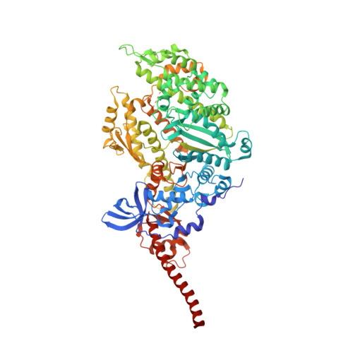
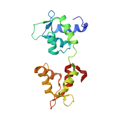
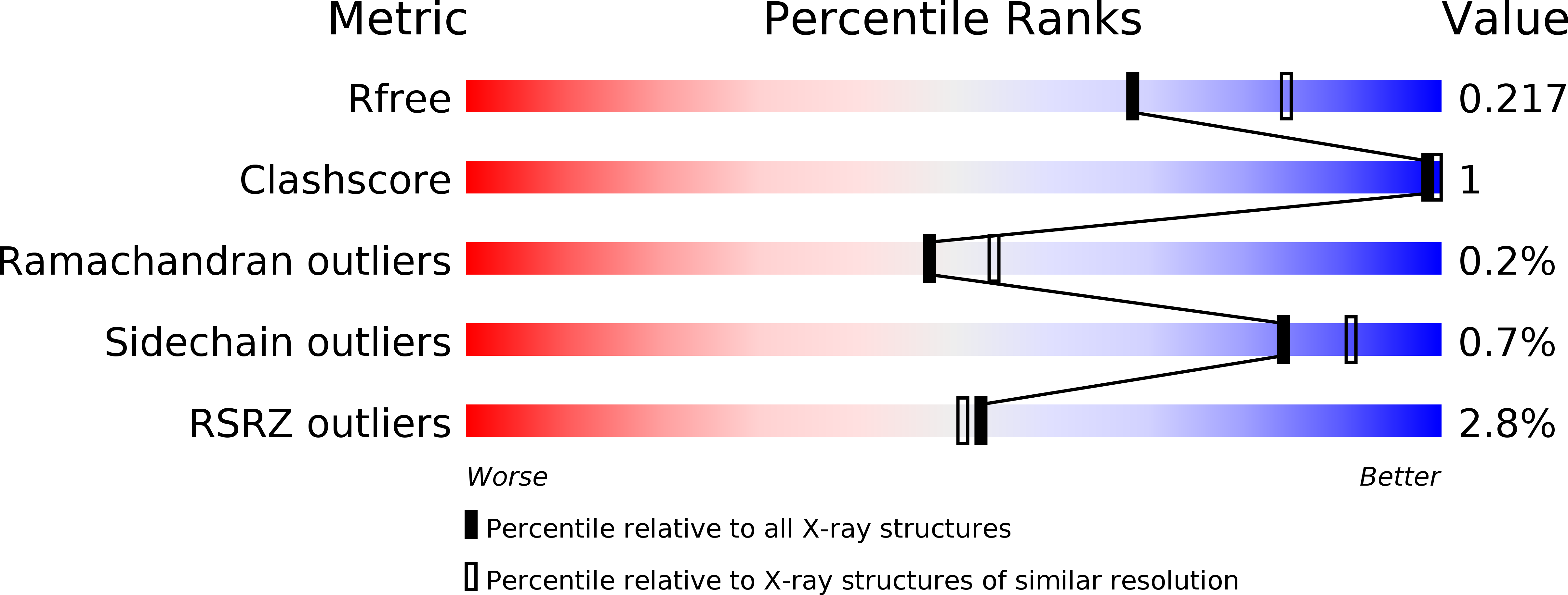
    Drosophila melanogaster is a powerful system for characterizing alternative myosin isoforms and modeling muscle diseases, but high-resolution structures of fruit fly contractile proteins have not been determined. Here we report the first x-ray crystal structure of an insect myosin: the D melanogaster skeletal muscle myosin II embryonic isoform (EMB). Using our system for recombinant expression of myosin heavy chain (MHC) proteins in whole transgenic flies, we prepared and crystallized stable proteolytic S1-like fragments containing the entire EMB motor domain bound to an essential light chain. We solved the x-ray crystal structure by molecular replacement and refined the resulting model against diffraction data to 2.2 Å resolution. The protein is captured in two slightly different renditions of the rigor-like conformation with a citrate of crystallization at the nucleotide binding site and exhibits structural features common to myosins of diverse classes from all kingdoms of life. All atom molecular dynamics simulations on EMB in its nucleotide-free state and a derivative homology model containing 61 amino acid substitutions unique to the indirect flight muscle isoform (IFI) suggest that differences in the identity of residues within the relay and the converter that are encoded for by MHC alternative exons 9 and 11, respectively, directly contribute to increased mobility of these regions in IFI relative to EMB. This suggests the possibility that alternative folding or conformational stability within these regions contribute to the observed functional differences in Drosophila EMB and IFI myosins.


  • Organizational Affiliation

    Structural Biochemistry Laboratory, Department of Chemistry & Biochemistry, San Diego State University, 5500 Campanile Drive, San Diego, CA 92182-1030, USA; Department of Biology and Molecular Biology Institute, San Diego State University, 5500 Campanile Drive, San Diego, CA 92182-4614, USA.


Macromolecules
Find similar proteins by:  (by identity cutoff)  |  3D Structure
Entity ID: 1
MoleculeChains Sequence LengthOrganismDetailsImage
Myosin heavy chain, muscle
A, C
810Drosophila melanogasterMutation(s): 0 
Gene Names: MhcCG17927
UniProt
Find proteins for P05661-9 (Drosophila melanogaster)
Explore P05661-9 
Go to UniProtKB:  P05661-9
Entity Groups  
Sequence Clusters30% Identity50% Identity70% Identity90% Identity95% Identity100% Identity
UniProt GroupP05661-9
Sequence Annotations
Expand
  • Reference Sequence
Find similar proteins by:  (by identity cutoff)  |  3D Structure
Entity ID: 2
MoleculeChains Sequence LengthOrganismDetailsImage
Myosin light chain alkali
B, D
155Drosophila melanogasterMutation(s): 0 
Gene Names: Mlc1MLC-ALKCG5596
UniProt
Find proteins for P06742-2 (Drosophila melanogaster)
Explore P06742-2 
Go to UniProtKB:  P06742-2
Entity Groups  
Sequence Clusters30% Identity50% Identity70% Identity90% Identity95% Identity100% Identity
UniProt GroupP06742-2
Sequence Annotations
Expand
  • Reference Sequence
Small Molecules
Ligands 3 Unique
IDChains Name / Formula / InChI Key2D Diagram3D Interactions
FLC
Query on FLC

Download Ideal Coordinates CCD File 
E [auth A],
K [auth C]
CITRATE ANION
C6 H5 O7
KRKNYBCHXYNGOX-UHFFFAOYSA-K
GOL
Query on GOL

Download Ideal Coordinates CCD File 
F [auth A],
G [auth A],
L [auth C],
M [auth C]
GLYCEROL
C3 H8 O3
PEDCQBHIVMGVHV-UHFFFAOYSA-N
EDO
Query on EDO

Download Ideal Coordinates CCD File 
H [auth A],
I [auth A],
J [auth A],
N [auth C]
1,2-ETHANEDIOL
C2 H6 O2
LYCAIKOWRPUZTN-UHFFFAOYSA-N
Experimental Data & Validation

Experimental Data

  • Method: X-RAY DIFFRACTION
  • Resolution: 2.23 Å
  • R-Value Free: 0.220 
  • R-Value Work: 0.178 
  • R-Value Observed: 0.180 
  • Space Group: P 21 21 21
Unit Cell:
Length ( Å )Angle ( ˚ )
a = 108.554α = 90
b = 148.582β = 90
c = 148.734γ = 90
Software Package:
Software NamePurpose
PHENIXrefinement
XDSdata reduction
XDSdata scaling
BALBESphasing

Structure Validation

View Full Validation Report


Entry History & Funding Information

Deposition Data


Funding OrganizationLocationGrant Number
National Institutes of Health/National Institute of General Medical Sciences (NIH/NIGMS)United StatesR01 GM032443

Revision History  (Full details and data files)

  • Version 1.0: 2018-06-06
    Type: Initial release
  • Version 1.1: 2019-12-25
    Changes: Database references
  • Version 1.2: 2020-01-01
    Changes: Author supporting evidence
  • Version 1.3: 2024-03-13
    Changes: Data collection, Database references