5VRH

Apolipoprotein N-acyltransferase C387S active site mutant


Experimental Data Snapshot

  • Method: X-RAY DIFFRACTION
  • Resolution: 2.14 Å
  • R-Value Free: 0.250 
  • R-Value Work: 0.207 
  • R-Value Observed: 0.209 

wwPDB Validation   3D Report Full Report


Ligand Structure Quality Assessment 


This is version 1.3 of the entry. See complete history


Literature

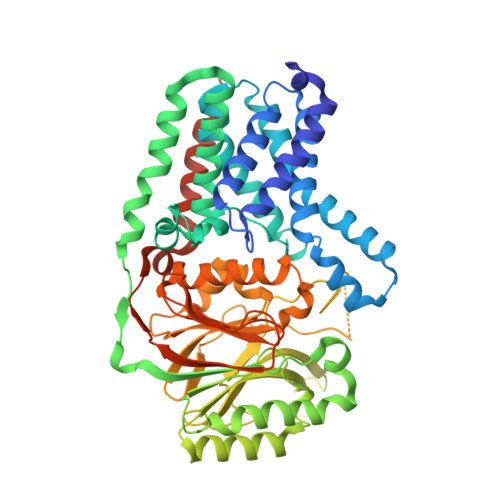
Structural insights into lipoprotein N-acylation by Escherichia coli apolipoprotein N-acyltransferase.

Noland, C.L.Kattke, M.D.Diao, J.Gloor, S.L.Pantua, H.Reichelt, M.Katakam, A.K.Yan, D.Kang, J.Zilberleyb, I.Xu, M.Kapadia, S.B.Murray, J.M.

(2017) Proc Natl Acad Sci U S A 114: E6044-E6053

  • DOI: https://doi.org/10.1073/pnas.1707813114
  • Primary Citation of Related Structures:  
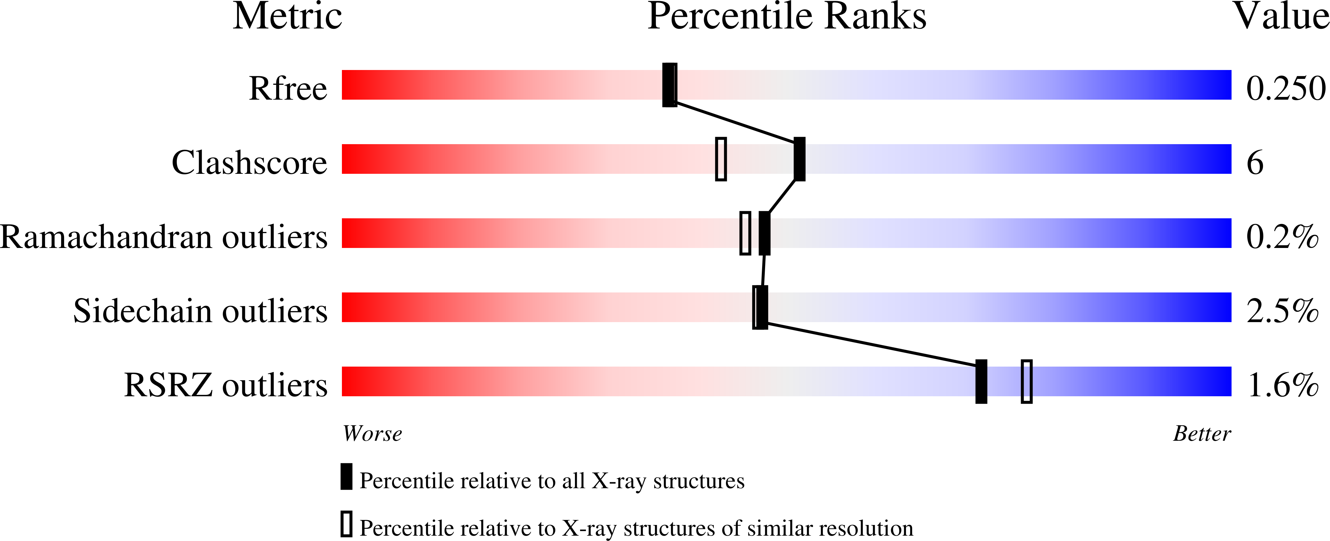
    5VRG, 5VRH

  • PubMed Abstract: 

    Gram-negative bacteria express a diverse array of lipoproteins that are essential for various aspects of cell growth and virulence, including nutrient uptake, signal transduction, adhesion, conjugation, sporulation, and outer membrane protein folding. Lipoprotein maturation requires the sequential activity of three enzymes that are embedded in the cytoplasmic membrane. First, phosphatidylglycerol:prolipoprotein diacylglyceryl transferase (Lgt) recognizes a conserved lipobox motif within the prolipoprotein signal sequence and catalyzes the addition of diacylglycerol to an invariant cysteine. The signal sequence is then cleaved by signal peptidase II (LspA) to give an N-terminal S-diacylglyceryl cysteine. Finally, apolipoprotein N -acyltransferase (Lnt) catalyzes the transfer of the sn -1-acyl chain of phosphatidylethanolamine to this N-terminal cysteine, generating a mature, triacylated lipoprotein. Although structural studies of Lgt and LspA have yielded significant mechanistic insights into this essential biosynthetic pathway, the structure of Lnt has remained elusive. Here, we present crystal structures of wild-type and an active-site mutant of Escherichia coli Lnt. The structures reveal a monomeric eight-transmembrane helix fold that supports a periplasmic carbon-nitrogen hydrolase domain containing a Cys-Glu-Lys catalytic triad. Two lipids are bound at the active site in the structures, and we propose a putative phosphate recognition site where a chloride ion is coordinated near the active site. Based on these structures and complementary cell-based, biochemical, and molecular dynamics approaches, we propose a mechanism for substrate engagement and catalysis by E. coli Lnt.


  • Organizational Affiliation

    Department of Structural Biology, Genentech, Inc., South San Francisco, CA 94080.


Macromolecules
Find similar proteins by:  (by identity cutoff)  |  3D Structure
Entity ID: 1
MoleculeChains Sequence LengthOrganismDetailsImage
Apolipoprotein N-acyltransferase522Escherichia coli K-12Mutation(s): 1 
Gene Names: lntcutEb0657JW0654
EC: 2.3.1
Membrane Entity: Yes 
UniProt
Find proteins for P23930 (Escherichia coli (strain K12))
Explore P23930 
Go to UniProtKB:  P23930
Entity Groups  
Sequence Clusters30% Identity50% Identity70% Identity90% Identity95% Identity100% Identity
UniProt GroupP23930
Sequence Annotations
Expand
  • Reference Sequence
Small Molecules
Ligands 5 Unique
IDChains Name / Formula / InChI Key2D Diagram3D Interactions
OLC
Query on OLC

Download Ideal Coordinates CCD File 
H [auth A],
I [auth A]
(2R)-2,3-dihydroxypropyl (9Z)-octadec-9-enoate
C21 H40 O4
RZRNAYUHWVFMIP-GDCKJWNLSA-N
OLB
Query on OLB

Download Ideal Coordinates CCD File 
B [auth A],
C [auth A]
(2S)-2,3-dihydroxypropyl (9Z)-octadec-9-enoate
C21 H40 O4
RZRNAYUHWVFMIP-QJRAZLAKSA-N
PLM
Query on PLM

Download Ideal Coordinates CCD File 
E [auth A],
F [auth A]
PALMITIC ACID
C16 H32 O2
IPCSVZSSVZVIGE-UHFFFAOYSA-N
CL
Query on CL

Download Ideal Coordinates CCD File 
G [auth A]CHLORIDE ION
Cl
VEXZGXHMUGYJMC-UHFFFAOYSA-M
MG
Query on MG

Download Ideal Coordinates CCD File 
D [auth A]MAGNESIUM ION
Mg
JLVVSXFLKOJNIY-UHFFFAOYSA-N
Experimental Data & Validation

Experimental Data

  • Method: X-RAY DIFFRACTION
  • Resolution: 2.14 Å
  • R-Value Free: 0.250 
  • R-Value Work: 0.207 
  • R-Value Observed: 0.209 
  • Space Group: P 1 21 1
Unit Cell:
Length ( Å )Angle ( ˚ )
a = 52.671α = 90
b = 72.586β = 101.73
c = 75.61γ = 90
Software Package:
Software NamePurpose
Aimlessdata scaling
PHASERphasing
PHENIXrefinement
PDB_EXTRACTdata extraction
XDSdata reduction

Structure Validation

View Full Validation Report


Ligand Structure Quality Assessment 


Entry History 

Deposition Data

Revision History  (Full details and data files)

  • Version 1.0: 2017-07-12
    Type: Initial release
  • Version 1.1: 2017-07-26
    Changes: Database references
  • Version 1.2: 2017-08-09
    Changes: Database references
  • Version 1.3: 2023-10-04
    Changes: Data collection, Database references, Refinement description