


Crystal Structure of a Hypothetical Protein from Neisseria gonorrhoeae with bound ppGpp
Dranow, D.M., Abendroth, J., Lorimer, D.D., Edwards, T.E.To be published.
Experimental Data Snapshot
Entity ID: 1 | |||||
|---|---|---|---|---|---|
| Molecule | Chains | Sequence Length | Organism | Details | Image |
| Putative phosphoribosyltransferase | 183 | Neisseria gonorrhoeae | Mutation(s): 0 Gene Names: ESCNG_40048, WHOF_00431, WHOF_02526C, WHOG_01129, WHOG_02374C, WHOK_01250, WHOK_02372C, WHOM_00300, WHOM_02380C, WHON_00453... | 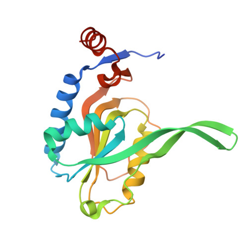 | |
UniProt | |||||
Find proteins for A0A1D3HIA2 (Neisseria gonorrhoeae) Explore A0A1D3HIA2 Go to UniProtKB: A0A1D3HIA2 | |||||
Entity Groups | |||||
| Sequence Clusters | 30% Identity50% Identity70% Identity90% Identity95% Identity100% Identity | ||||
| UniProt Group | A0A1D3HIA2 | ||||
Sequence AnnotationsExpand | |||||
| |||||
| Ligands 3 Unique | |||||
|---|---|---|---|---|---|
| ID | Chains | Name / Formula / InChI Key | 2D Diagram | 3D Interactions | |
| G4P Query on G4P | D [auth A] | GUANOSINE-5',3'-TETRAPHOSPHATE C10 H17 N5 O17 P4 BUFLLCUFNHESEH-UUOKFMHZSA-N |  | ||
| EDO Query on EDO | B [auth A], C [auth A] | 1,2-ETHANEDIOL C2 H6 O2 LYCAIKOWRPUZTN-UHFFFAOYSA-N |  | ||
| MG Query on MG | E [auth A], F [auth A] | MAGNESIUM ION Mg JLVVSXFLKOJNIY-UHFFFAOYSA-N |  | ||
| Length ( Å ) | Angle ( ˚ ) |
|---|---|
| a = 81.27 | α = 90 |
| b = 103.93 | β = 90 |
| c = 105.54 | γ = 90 |
| Software Name | Purpose |
|---|---|
| XSCALE | data scaling |
| PHENIX | refinement |
| PDB_EXTRACT | data extraction |
| XDS | data reduction |
| MoRDa | phasing |
RCSB PDB (citation) is hosted by
RCSB PDB is a member of the

