5VN1

horse liver alcohol dehydrogenae complexed with NADH (R,S)-N-1-methylhexylformamide

  • Classification: OXIDOREDUCTASE
  • Organism(s): Equus caballus
  • Mutation(s): No 

  • Deposited: 2017-04-28 Released: 2017-05-10 
  • Deposition Author(s): Plapp, B.V., Ramaswamy, S., Ferraro, D.J., Baskar Raj, S.
  • Funding Organization(s): National Institutes of Health/National Institute on Alcohol Abuse and Alcoholism (NIH/NIAAA), National Institutes of Health/National Institute of General Medical Sciences (NIH/NIGMS)

Experimental Data Snapshot

  • Method: X-RAY DIFFRACTION
  • Resolution: 1.25 Å
  • R-Value Free: 0.187 
  • R-Value Work: 0.151 
  • R-Value Observed: 0.152 

wwPDB Validation   3D Report Full Report


Ligand Structure Quality Assessment 


This is version 1.4 of the entry. See complete history


Literature

Horse Liver Alcohol Dehydrogenase: Zinc Coordination and Catalysis.

Plapp, B.V.Savarimuthu, B.R.Ferraro, D.J.Rubach, J.K.Brown, E.N.Ramaswamy, S.

(2017) Biochemistry 56: 3632-3646

  • DOI: https://doi.org/10.1021/acs.biochem.7b00446
  • Primary Citation of Related Structures:  
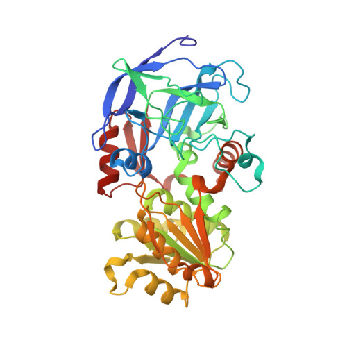
    5VJ5, 5VJG, 5VKR, 5VL0, 5VN1

  • PubMed Abstract: 

    During catalysis by liver alcohol dehydrogenase (ADH), a water bound to the catalytic zinc is replaced by the oxygen of the substrates. The mechanism might involve a pentacoordinated zinc or a double-displacement reaction with participation by a nearby glutamate residue, as suggested by studies of human ADH3, yeast ADH1, and some other tetrameric ADHs. Zinc coordination and participation of water in the enzyme mechanism were investigated by X-ray crystallography. The apoenzyme and its complex with adenosine 5'-diphosphoribose have an open protein conformation with the catalytic zinc in one position, tetracoordinated by Cys-46, His-67, Cys-174, and a water molecule. The bidentate chelators 2,2'-bipyridine and 1,10-phenanthroline displace the water and form a pentacoordinated zinc. The enzyme-NADH complex has a closed conformation similar to that of ternary complexes with coenzyme and substrate analogues; the coordination of the catalytic zinc is similar to that found in the apoenzyme, except that a minor, alternative position for the catalytic zinc is ∼1.3 Å from the major position and closer to Glu-68, which could form the alternative coordination to the catalytic zinc. Complexes with NADH and N-1-methylhexylformamide or N-benzylformamide (or with NAD + and fluoro alcohols) have the classical tetracoordinated zinc, and no water is bound to the zinc or the nicotinamide rings. The major forms of the enzyme in the mechanism have a tetracoordinated zinc, where the carboxylate group of Glu-68 could participate in the exchange of water and substrates on the zinc. Hydride transfer in the Michaelis complexes does not involve a nearby water.


  • Organizational Affiliation

    Department of Biochemistry, The University of Iowa , Iowa City, Iowa 52242, United States.


Macromolecules
Find similar proteins by:  (by identity cutoff)  |  3D Structure
Entity ID: 1
MoleculeChains Sequence LengthOrganismDetailsImage
Alcohol dehydrogenase E chain
A, B, C, D
374Equus caballusMutation(s): 0 
EC: 1.1.1.1
UniProt
Find proteins for P00327 (Equus caballus)
Explore P00327 
Go to UniProtKB:  P00327
Entity Groups  
Sequence Clusters30% Identity50% Identity70% Identity90% Identity95% Identity100% Identity
UniProt GroupP00327
Sequence Annotations
Expand
  • Reference Sequence
Small Molecules
Ligands 5 Unique
IDChains Name / Formula / InChI Key2D Diagram3D Interactions
NAI
Query on NAI

Download Ideal Coordinates CCD File 
G [auth A],
K [auth B],
P [auth C],
V [auth D]
1,4-DIHYDRONICOTINAMIDE ADENINE DINUCLEOTIDE
C21 H29 N7 O14 P2
BOPGDPNILDQYTO-NNYOXOHSSA-N
NMH
Query on NMH

Download Ideal Coordinates CCD File 
R [auth C](R)-N-(1-METHYL-HEXYL)-FORMAMIDE
C8 H17 N O
GFVRKPKAQHTAQK-MRVPVSSYSA-N
NWH
Query on NWH

Download Ideal Coordinates CCD File 
H [auth A],
L [auth B],
Q [auth C],
W [auth D]
N-[(2S)-heptan-2-yl]formamide
C8 H17 N O
GFVRKPKAQHTAQK-QMMMGPOBSA-N
MRD
Query on MRD

Download Ideal Coordinates CCD File 
M [auth B],
S [auth C]
(4R)-2-METHYLPENTANE-2,4-DIOL
C6 H14 O2
SVTBMSDMJJWYQN-RXMQYKEDSA-N
ZN
Query on ZN

Download Ideal Coordinates CCD File 
E [auth A]
F [auth A]
I [auth B]
J [auth B]
N [auth C]
E [auth A],
F [auth A],
I [auth B],
J [auth B],
N [auth C],
O [auth C],
T [auth D],
U [auth D]
ZINC ION
Zn
PTFCDOFLOPIGGS-UHFFFAOYSA-N
Binding Affinity Annotations 
IDSourceBinding Affinity
NMH Binding MOAD:  5VN1 Ki: 5400 (nM) from 1 assay(s)
NWH Binding MOAD:  5VN1 Ki: 5400 (nM) from 1 assay(s)
Experimental Data & Validation

Experimental Data

  • Method: X-RAY DIFFRACTION
  • Resolution: 1.25 Å
  • R-Value Free: 0.187 
  • R-Value Work: 0.151 
  • R-Value Observed: 0.152 
  • Space Group: P 1 21 1
Unit Cell:
Length ( Å )Angle ( ˚ )
a = 50.212α = 90
b = 180.808β = 106.12
c = 86.841γ = 90
Software Package:
Software NamePurpose
XSCALEdata scaling
REFMACrefinement
PDB_EXTRACTdata extraction
XDSdata reduction
X-PLORphasing
Omodel building

Structure Validation

View Full Validation Report


Ligand Structure Quality Assessment 


Entry History & Funding Information

Deposition Data


Funding OrganizationLocationGrant Number
National Institutes of Health/National Institute on Alcohol Abuse and Alcoholism (NIH/NIAAA)United StatesAA00279
National Institutes of Health/National Institute of General Medical Sciences (NIH/NIGMS)United StatesT32 GM08365

Revision History  (Full details and data files)

  • Version 1.0: 2017-05-10
    Type: Initial release
  • Version 1.1: 2017-07-05
    Changes: Database references
  • Version 1.2: 2017-08-02
    Changes: Author supporting evidence, Database references
  • Version 1.3: 2019-12-11
    Changes: Author supporting evidence
  • Version 1.4: 2023-10-04
    Changes: Data collection, Database references, Derived calculations, Refinement description