5VDK

Crystal structure of human WEE2 kinase domain in complex with MK1775


Experimental Data Snapshot

  • Method: X-RAY DIFFRACTION
  • Resolution: 2.70 Å
  • R-Value Free: 0.284 
  • R-Value Work: 0.225 
  • R-Value Observed: 0.228 

wwPDB Validation   3D Report Full Report


Ligand Structure Quality Assessment 


This is version 1.3 of the entry. See complete history


Literature

Structural Basis of Wee Kinases Functionality and Inactivation by Diverse Small Molecule Inhibitors.

Zhu, J.Y.Cuellar, R.A.Berndt, N.Lee, H.E.Olesen, S.H.Martin, M.P.Jensen, J.T.Georg, G.I.Schonbrunn, E.

(2017) J Med Chem 60: 7863-7875

  • DOI: https://doi.org/10.1021/acs.jmedchem.7b00996
  • Primary Citation of Related Structures:  
    5V5Y, 5VC3, 5VC4, 5VC5, 5VC6, 5VCV, 5VCW, 5VCX, 5VCY, 5VCZ, 5VD0, 5VD1, 5VD2, 5VD3, 5VDK

  • PubMed Abstract: 

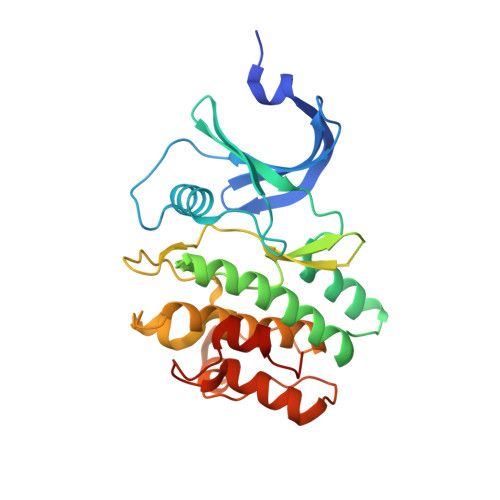
    Members of the Wee family of kinases negatively regulate the cell cycle via phosphorylation of CDK1 and are considered potential drug targets. Herein, we investigated the structure-function relationship of human Wee1, Wee2, and Myt1 (PKMYT1). Purified recombinant full-length proteins and kinase domain constructs differed substantially in phosphorylation states and catalytic competency, suggesting complex mechanisms of activation. A series of crystal structures reveal unique features that distinguish Wee1 and Wee2 from Myt1 and establish the structural basis of differential inhibition by the widely used Wee1 inhibitor MK-1775. Kinome profiling and cellular studies demonstrate that, in addition to Wee1 and Wee2, MK-1775 is an equally potent inhibitor of the polo-like kinase PLK1. Several previously unrecognized inhibitors of Wee kinases were discovered and characterized. Combined, the data provide a comprehensive view on the catalytic and structural properties of Wee kinases and a framework for the rational design of novel inhibitors thereof.


  • Organizational Affiliation

    Drug Discovery Department, Moffitt Cancer Center , Tampa, Florida 33612, United States.


Macromolecules
Find similar proteins by:  (by identity cutoff)  |  3D Structure
Entity ID: 1
MoleculeChains Sequence LengthOrganismDetailsImage
Wee1-like protein kinase 2297Homo sapiensMutation(s): 0 
Gene Names: WEE2WEE1B
EC: 2.7.10.2
UniProt & NIH Common Fund Data Resources
Find proteins for P0C1S8 (Homo sapiens)
Explore P0C1S8 
Go to UniProtKB:  P0C1S8
PHAROS:  P0C1S8
GTEx:  ENSG00000214102 
Entity Groups  
Sequence Clusters30% Identity50% Identity70% Identity90% Identity95% Identity100% Identity
UniProt GroupP0C1S8
Sequence Annotations
Expand
  • Reference Sequence
Small Molecules
Ligands 2 Unique
IDChains Name / Formula / InChI Key2D Diagram3D Interactions
8X7
Query on 8X7

Download Ideal Coordinates CCD File 
B [auth A]1-[6-(2-hydroxypropan-2-yl)pyridin-2-yl]-6-{[4-(4-methylpiperazin-1-yl)phenyl]amino}-2-(prop-2-en-1-yl)-1,2-dihydro-3H-pyrazolo[3,4-d]pyrimidin-3-one
C27 H32 N8 O2
BKWJAKQVGHWELA-UHFFFAOYSA-N
PO4
Query on PO4

Download Ideal Coordinates CCD File 
C [auth A],
D [auth A],
E [auth A]
PHOSPHATE ION
O4 P
NBIIXXVUZAFLBC-UHFFFAOYSA-K
Binding Affinity Annotations 
IDSourceBinding Affinity
8X7 BindingDB:  5VDK Kd: min: 3.9, max: 27 (nM) from 2 assay(s)
Experimental Data & Validation

Experimental Data

  • Method: X-RAY DIFFRACTION
  • Resolution: 2.70 Å
  • R-Value Free: 0.284 
  • R-Value Work: 0.225 
  • R-Value Observed: 0.228 
  • Space Group: P 32
Unit Cell:
Length ( Å )Angle ( ˚ )
a = 68.81α = 90
b = 68.81β = 90
c = 58.04γ = 120
Software Package:
Software NamePurpose
PHENIXrefinement
XSCALEdata scaling
PDB_EXTRACTdata extraction
XDSdata reduction
PHASERphasing

Structure Validation

View Full Validation Report


Ligand Structure Quality Assessment 


Entry History & Funding Information

Deposition Data


Funding OrganizationLocationGrant Number
National Institutes of Health/Eunice Kennedy Shriver National Institute of Child Health & Human Development (NIH/NICHD)United StatesUO1 HD076542

Revision History  (Full details and data files)

  • Version 1.0: 2017-08-23
    Type: Initial release
  • Version 1.1: 2017-10-11
    Changes: Database references
  • Version 1.2: 2019-12-11
    Changes: Author supporting evidence
  • Version 1.3: 2023-10-04
    Changes: Data collection, Database references, Refinement description