


Crystal structure of a serine hydroxymethyltransferase from Helicobacter pylori
Edwards, T.E., Mayclin, S.J., Lorimer, D.D., Seattle Structural Genomics Center for Infectious Disease (SSGCID)To be published.
Experimental Data Snapshot
wwPDB Validation 3D Report Full Report

Entity ID: 1 | |||||
|---|---|---|---|---|---|
| Molecule | Chains | Sequence Length | Organism | Details | Image |
| Serine hydroxymethyltransferase | 424 | Helicobacter pylori G27 | Mutation(s): 0 Gene Names: glyA, HPG27_169 EC: 2.1.2.1 | 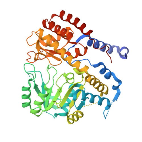 | |
UniProt | |||||
Find proteins for B5Z9V7 (Helicobacter pylori (strain G27)) Explore B5Z9V7 Go to UniProtKB: B5Z9V7 | |||||
Entity Groups | |||||
| Sequence Clusters | 30% Identity50% Identity70% Identity90% Identity95% Identity100% Identity | ||||
| UniProt Group | B5Z9V7 | ||||
Sequence AnnotationsExpand | |||||
| |||||
| Length ( Å ) | Angle ( ˚ ) |
|---|---|
| a = 57.97 | α = 90 |
| b = 91.02 | β = 90 |
| c = 161.32 | γ = 90 |
| Software Name | Purpose |
|---|---|
| XSCALE | data scaling |
| MOLREP | phasing |
| PHENIX | refinement |
| PDB_EXTRACT | data extraction |
| XDS | data reduction |
RCSB PDB (citation) is hosted by
RCSB PDB is a member of the

